

 PDF
PDF ePub
ePub Citation
 Print
Print


INTRODUCTION
Hernia of the umbilical cord is a rare condition that results from a failure in the development of the body wall folds in the human embryo. The hernia can contain small intestine, large intestine, liver, gall bladder, spleen or stomach. Segmental dilatation of the ileum (SDI) can be associated with hernia of the umbilical cord and requires resection during the surgery for abdominal wall repair.
CASE REPORT
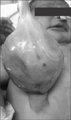
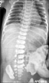
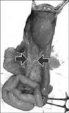
A full term 2.5 kg male baby was referred soon after birth as a case of omphalocele. The neonate had passed urine and small amount of meconium. The cry, tone and activity of the baby was good. On abdominal examination, there was a large sac 6×5 cm with a narrow pedicle of 2 cm (Fig. 1). The nasogastric tube was draining bile. An abdominal radiograph revealed few bowel loops inside the sac (Fig. 2). The baby was taken up for surgery after adequate resuscitation. Intra-operatively, the sac contained few loops of small intestine. There was a fusiform SDI, about 25 cm proximal to the ileocecal valve. Note was made of conspicuous mesenteric vessels or mesenteric folds in a V-shaped pattern towards the two ends of the SDI (Fig. 3). Resection and end to end anastomosis was done along with the abdominal wall repair. The child had an uneventful recovery and was discharged on postoperative day 7. The histopathology of the segment revealed normal ganglion cells and hypertrophy of muscle fibres.
DISCUSSION
Hernia of the umbilical cord is a type of omphalocele, where the diameter of the abdominal defect is less than 4 cm. The origin of the defect is a failure of the bowel to return to the body cavity from its physiological herniation during the 6th to 10th weeks of gestation. It is a rare disease with an incidence of about 2.5/10,000 births. Due to its association with chromosomal anomalies, cardiac defects and neural tube defects, it has a high rate of mortality of 25%. The herniated viscus is most commonly the small intestine which is covered by an amniotic membrane.
SDI is an even rarer entity with only around 150 cases reported in literature so far [1]. The first report was published by Swenson and Rathauser [2] in 1959 and detailed 3 cases involving the colon. The following criteria have thus been enumerated in order to diagnose segmental dilatation of the intestine; (i) limited bowel dilatation with a 3- to 4-fold increase in size, (ii) an abrupt transition between dilated and normal bowel, (iii) no intrinsic or extrinsic barrier distal to the dilatation, (iv) clinical picture of intestinal occlusion or sub-occlusion, (v) a normal neuronal plexus, and (vi) complete recovery after resection of the affected segment.
The coexistence of hernia of the umbilical cord and SDI is expectedly uncommon with less than 20 cases reported so far. At the outset, the etiology of SDI in a hernial sac appears to be compression at the umbilical ring, however this does not explain segmental dilatation in the colon, which does not herniate out from the abdominal wall, and hence cannot get strangulated at the umbilical ring.
Heller et al. [3] have proposed an elegant theory which if applied, implicates segmental dilatation to be the primary pathology. Inability of the bulbous intestine to return to the abdominal cavity results in the formation of a small omphalocele or hernia of the umbilical cord. The theory suggests that SDI, duplication cysts and diverticuli occur due to incomplete splitting of the notochord from the endoderm during the 3rd week of development. Remnants of the primitive gut form a duplication cyst with or without ectopic tissue, at the mesenteric border of the intestine which eventually gets resorbed with the adjoining normal intestine forming a grossly dilated segment. The dilatation and haphazard arrangement of muscle fibres cause the functional obstruction at the segment.
The presence of mesenteric folds, abnormal mesenteric vessels demarcating the two ends of the dilated segment also suggest the presence of an erstwhile duplicated segment with its blood supply, which eventually got resorbed. This abnormal folds/vessel in the mesentery have been noted by some authors and is evident on some of the photographs in previous published articles [45].
SDI can be associated with vertebral anomalies and ectopic tissue, another marker of its origins from the notochord. SDI can also be associated with malrotation, Meckel's diverticulum, intestinal atresia, spina bifida and bladder exstrophy [6]. In our case, however, there were no associated anomalies.
Of the 16 previous reports of SDI associated with omphalocele minor, 14 were present in neonates [67]. One was diagnosed with obstruction 1 year after omphalocele surgery and another, 8 years after surgery. These patients who presented later and needed reoperation were the patients in whom the dilated segment was not excised during the first surgery. Diversion of SDI has also been attempted which can cause anemia, hypoproteinemia and severe malnutrition in the patient, necessitating another surgery for the removal of the segment. The standard of care in a patient with hernia of umbilical cord and SDI is abdominal wall repair along with resection of dilated segment and end to end anastomosis in the same sitting.
 XML Download