

 PDF
PDF ePub
ePub Citation
 Print
Print


INTRODUCTION
Gastrointestinal tract duplications are rare conditions, comprising 0.1% to 0.3% of all congenital anomalies. Duplications can occur throughout the gastrointestinal tract, from the mouth to the anus [12]. Anorectal duplications account for only 5% of all duplications and are primarily found in the retro-rectal space. Anterior anorectal duplications are extremely rare [3]. Anterior anorectal duplications are tubular duplications connected to the colon and open like a distal fistula, but are located anterior to the rectum and behind the bladder and vagina. It is challenging to diagnose anterior anorectal duplication, both because of its rarity and because it can present in different ways. Perirectal abscess, fistula-in-ano, recurrent fistulae due to Crohn's disease, infected dermoid cysts, sacrococcygeal teratomas, and anterior sacral meningoceles must be considered part of the differential diagnosis [34]. Here, we present the case of an anterior anorecto-sigmoid colonic tubular duplication initially presenting as a rectovaginal fistula in a 2-month-old infant. We also provide a brief literature review.
CASE REPORT
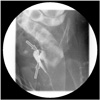
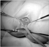
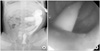
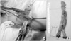
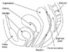
A 2-month-old female infant presented to our hospital with a 1-month history of stool passing through the vagina. She was born at 36 weeks' gestation weighing 1.875 kg and was the second infant of a twin. She had a ventricular septal defect that closed spontaneously at 1 month of age. Initially, she was diagnosed with a rectovaginal fistula. Despite conservative therapy, including a sitz bath, for 5 months, there was no improvement in her symptoms, and most of the stool still appeared to pass through the vagina. A colon study indicated a rectovaginal fistula directly connected to the distal rectum (Fig. 1). Under general anesthesia, a small opening located posterior to and near the vagina at vestibule was found. The structure that had been misdiagnosed as a fistula based on the colon study was not present. The patient had a normally sited anus, an anterior ectopic anus, and a double vagina (Fig. 2). A catheter was inserted through the ectopic anus, and an intraoperative contrast study and sigmoidoscopy were performed. A parallel tubular duplication with a common wall was found. The common wall extended 5 cm from the anal verge to the sigmoid colon and then 20 cm to the level of the proximal sigmoid colon. Proximally, the anterior duplication and normal colon merged and formed a normal colon. Because most of the stool moved through the anterior duplication and because the diameter of the anterior duplication was larger than that of the normally located colon (Fig. 3), complete resection of both the anterior duplicated bowel and posterior native bowel up to the merging point with colo-anal anastomosis was performed (Fig. 4). Fig. 5 shows a schematic diagram of surgical findings. Pathologic examination confirmed a duplicated colonic segment with a common proper muscle layer. The patient was discharged on the 8th postoperative day without incident. During a 3-year follow-up, she defecated normally without complications.
DISCUSSION
Gastrointestinal tract duplications occur with an incidence of 1 in 4,000 to 5,000 live births. The incidence of anorectal and colonic duplications has been reported as 3% to 8% and 4% to 18% of all duplications, respectively [356]. Anorectal duplications comprise a small proportion of enteric duplications, with only 70 cases reported to date in the literature. Based on their embryogenesis, anorectal duplications would be expected to be located posterior to the rectum. In fact, most anorectal duplications are located posterior to the anus; anterior rectal duplications are very rare, with fewer than 10 cases reported [7].
Although the causes of alimentary tract duplication are uncertain, many theories have been proposed to explain their pathophysiology, including the abortive twinning theory, persistent embryonic diverticular theory, split notochord theory, and aberrant luminal recanalization theory. However, no single theory can explain all forms of intestinal duplication [268]. Anorectal duplications are generally attributed to a simple “pinching off” of the diverticulum in the 8th to 9th week of gestation or to more complex “caudal twinning” occurring when the embryo is 10 mm in length during the 5th week of gestation [3].
Symptoms and findings depend on the location of the duplication [2]. Most patients with anorectal duplications are asymptomatic. The most frequent symptoms include progressive constipation, prolapse of the anorectal mucosa, and mucosal discharge. More severe symptoms have also been reported, including recurrent fistulae, pain, or infection in the duplicated cyst, rectal bleeding from ulceration, or ectopic gastric mucosa. The severity of these symptoms increases with the age at presentation. Malignant degeneration of duplications has also been reported [3910]. The symptoms reported for anterior anorectal duplications are similar to those for posterior anorectal duplications, including recurrent gastroenteritis and failure to thrive, isolated rectal bleeding, chronic constipation with soiling, and rectal prolapse [7]. Due to the differing presentations for anterior anorectal duplications, various diseases must be considered part of the differential diagnosis, including sacrococcygeal teratoma, anal fistula due to Crohn's disease, rectal prolapse, dermoid cysts, hydrocolpos, chordoma, leiomyosarcoma, and hydrometrocolpos [347]. Because it is challenging to distinguish anterior anorectal duplications from other diseases, delayed diagnosis can occur[4].
Duplications of the hindgut, such as anorectal duplications, are frequently accompanied by genitourinary tract anomalies and have complicated structures; thus, they tend to be challenging and complex. When encountering such duplications, genitourinary tract involvement should be considered [2].
Various diagnostic modalities can assist in diagnosis, such as abdominal ultrasonography, contrast studies, computed tomography, and MRI [7]. In particular, MRI can provide multiplanar views, allowing identification of pelvic structures and ruling out other diagnoses [67]. To evaluate communication between duplications and normal bowels, contrast studies can be helpful in accurately delineating the anatomy of the duplicated rectum [6]. In the present case, we performed a preoperative contrast study, but we initially misinterpreted the contrast spill as a fistula. We reached a final diagnosis intraoperatively after re-performing the contrast study. The second contrast study clearly revealed the anterior anorectocolonic tubular duplication.
Total surgical excision of all duplications is recommended because of the potential for long-term complications. If an anorectal duplication is not treated, secretions accumulate in the lumen of the duplication, leading to infection with age [1]. Malignant changes in anorectal duplications have also been reported in adult patients, although not in pediatric populations [110]. When there was no evidence of fistula formation between the duplication cyst and vagina, cyst unroofing and residual mucosa ablation was applied [7]. In the other case of anterior rectal duplication without rectovaginal fistula, septum division via surgical staplers was performed [3].
The location of the duplication determines the direction of approach. Perineal or posterior sagittal approaches in the prone position have primarily been used in cases of posterior rectal duplication and anal canal duplication [1], whereas abdominoperineal approaches have been chosen for anterior rectal duplications [37]. One reported case of posterior anorectal duplication extending to the colon was initially treated with a posterior sagittal approach, but as the dissection proceeded cephalad, it became difficult to manage, and laparotomy was also performed [6]. In our case, a perineal approach was first attempted because the lesion was misdiagnosed as a rectovaginal fistula; however, when the anterior anorectosigmoid duplication was found, the operation proceeded using an abdominal approach.
In summary, for infants who present with stool discharging from the vagina, anterior anorectal duplication should be considered as a differential diagnosis, despite its rarity. Because it is challenging to diagnose, careful evaluation is required.
 XML Download