

 PDF
PDF ePub
ePub Citation
 Print
Print


INTRODUCTION
Soft tissue hemangioendothelioma (STHE) is a rare vascular tumor, characterized by an increase in the blood vessel endothelial cells of soft tissue. Its prognosis lies between that of benign hemangioma and malignant angiosarcoma [1].
The most common presentation of STHE is a palpable mass. Surgical treatment has been recommended for STHE because it displays the characteristics of borderline malignancy. The prognosis of STHE depends on the tumor location, size, resection margin, pathologic subtype, and grade [123].
The prognosis of STHE for children in poorly understood, due to its rarity in this age group. In this study, we review the clinical characteristics of 8 pediatric cases and the results of their treatment, and we discuss prognostic factors regarding STHE in children.
METHODS
We retrospectively analyzed data from the medical records of 8 patients with STHE, who were diagnosed at Seoul National University Hospital through histopathological examination between January 1995 and June 2015. We evaluated the clinical presentation, diagnosis,treatment, and post-treatment status for each case. Soft tissue includes connective tissues such as tendons, ligaments, fascia, skin, fibrous tissues, fat, and synovial membranes, as well as non-connective tissues such as muscles, nerves, and blood vessels [1]. We excluded hemangioendothelioma (HE) of the liver, lungs, and pancreas.
We categorized surgical resection into three subtypes; microscopic complete resection (R0), macroscopic complete resection (R1) (in which complete resection was conducted macroscopically, but HE cells were confirmed microscopically in the resection margins), and macroscopic incomplete resection (R2).
The median duration of patient monitoring was 72 months (range, 4-165 months). Current patient status was divided into five categories; no evidence of disease, remission, no progression, recurrence, and regrowth. Remission referred to cases in which the patient showed no HE-related symptoms after R1. No progression referred to cases where remnant tumors showed no signs of regrowth, and the patient did not experience any additional symptoms after R2.
We conducted this study in compliance with the principles of the Declaration of Helsinki. This study was reviewed and approved by the Institutional Review Board of Seoul National University Hospital (IRB no. 1207-082-418). The informed consent was waived.
RESULTS
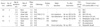
Of the 8 patients, 5 were male and 3 were female. The median age at the time of surgery was 1.2 years (range, 3 months to 12.2 years). The clinical presentation of 6 of the patients was a palpable tumor, and the other 2 patients presented with facial paralysis. The following tumor locations were observed head (2 patients), neck (2 patients), chest wall (1 patient), sacrococcyx (1 patient), upper limb (1 patient), and lower limb (1 patient). Radiologic evaluation was performed before surgery using one or more of the following techniques: ultrasonography (2 patients), CT (4 patients), and MRI (5 patients). The imaging results led to suspected diagnoses of hemangioma (4 patients), schwannoma (1 patient), teratoma (1 patient), desmoid tumor (1 patient), and an unidentifiable malignant tumor (1 patient). The median tumor size was 4.0 cm (range, 2.5-8.0 cm). Incisional biopsy was performed before surgery in 3 patients, and the pathologic results suggested hemangioma, histiocytoma and HE, respectively. All of the patients in this study showed normal platelet counts and coagulation profiles, with no accompanying Kasabach-Merritt syndrome [4].
The initial treatment for all 8 patients was surgical resection (Table 1). Of these, 3 patients underwent microscopic complete resection (R0), 2 patients underwent macroscopic complete resection (R1), and 3 patients underwent macroscopic incomplete resection (R2). The R2 resection tended to be performed for head and neck lesions, particularly if the lesion was large, and adjacent to major vessels and cranial nerves. Histopathological examination after surgery confirmed kaposiform HE in 6 patients, retiform HE in 1 patient, andepithelioid HE in 1 patient. Tumor grade 2 was confirmed in 2 patients, according to the French Federation Nationale des Centers de LutteContre le Cancer (FNCLCC) grading system [5].
After surgical resection, the main sequelae were gait disturbance, which occurred in the patient with STHE of the lower limb (R1), and vocal cord palsy and hearing impairment, which occurred in patients with STHE of the head (R2).
Four patients (R1, 2 patients; R2, 2 patients) were treated with adjuvant IFN therapy. Additionally, 4 patients underwent second surgery (R0, 1 patient; R1, 2 patients; and R2, 1 patient). Three patients received IFN therapy due to recurrence or regrowth, including 2 patients who were diagnosed as FNCLCC grade 2. One patient, who underwent R2 resection for a head lesion, underwent resection 3 more times, but died 11 months after the first surgery, due to local progression. Currently, 6 patients are doing well, without recurrence. However, distant metastasis to the scalp was observed in 1 patient withepithelioid HE of the sacrococcyx (FNCLCC grade 2), despite reoperation and IFN therapy. The scalp mass was excised and the patient is currently receiving oral cyclophosphamide therapy.
DISCUSSION
Until now, it has been difficult to diagnose and predict the degree of malignancy of STHE, before surgery [67]. Even though radiologic studies (ultrasonography, CT, and MRI) and histopathological evaluation (fine-needle aspiration cytology and incisional biopsy) could be helpful in the diagnosis of STHE, previous studies have shown that it is difficult to diagnose STHE accurately using fine-needle aspiration cytology [89]. When the tumor is not thought to be simple hemangioma, incisional biopsy may provide an exact diagnosis and an estimation of the malignancy grade [1]. However, for pediatric patients, incisional biopsy is limited by its high false-negative rate and the need for general anesthesia. In the present study, no patients were accurately diagnosed by preoperative radiologic examination, and only 1 of the 3 patients who had an incisional biopsy was diagnosed with HE accurately. Therefore, either incisional or excisional biopsies must also be performed to achieve accurate diagnosis in pediatric patients.
The primary treatment of STHE is surgical resection. R0 is usually recommended, because STHE is not a benign tumor in most cases. If R0 resection is not achieved, there may be a high risk of recurrence and metastasis, which could decrease overall survival rate [31011].
However, it is debatable whether extensive radical surgery should be conducted, like in cases of angiosarcoma [310]. The prognosis of STHE differs from that of malignant vascular tumors, and even high-risk epithelioid HEs have better prognoses than angiosarcomas [31012]. In this study, some patients achieved a stable state after partial resection (even R2). In addition, 3 patients experienced major sequelae after surgery, such as gait disturbance, hearing impairment, and vocal cord palsy. Therefore, function-preserving surgery seems advisable when radical resection could lead to major side effects.
Although chemotherapy would be helpful in treating STHE with Kasabach-Merritt syndrome, the scope of chemotherapy in treating STHE alone remains unclear. [11131415]. Only a few previous studies have shown the benefits of adjuvant chemotherapy, and the results were highly estimated. In these reports, either combination therapy or mono-therapy was used, consisting of steroids, IFNs, mechanistic target of rapamycin (mTOR) inhibitors, and/or chemotherapeutic agents such as vincristine [36]. In the present study, 3 of the 4 patients who underwent IFN chemotherapy after surgery eventually showed regrowth and/or distant metastasis. We suggest the need for further research on the role of IFN therapy for treating STHE.
There are generally three isoforms of HE; an intermediate-locally aggressive isoform (kaposiform), an intermediate-rarely metastasizing isoform (retiform/composite), and a malignant isoform (epithelioid) [12]. Pathologic isoforms exhibit differences in malignancy, recurrence, metastasis, and prognosis. Kaposiform HE may have a prognosis that lies between a benign and malignant tumor, because it can re-grow locally, but usually does not metastasize. Using immunohistochemistry, HE isoforms can be differentiated from other endothelial tumors, such as angiosarcoma and tufted angioma[6]. For example, the expression of CD34 and CD31 indicates kaposiform HE, and the expression of CD31, Friend leukemia integration 1 transcription factor (FLI-1), and erythroblast transformation-specific (ETS)-related gene (ERG), indicates epitheloid HE [610].
The prognosis for kaposiform HE accompanied by Kasabach-Merritt syndrome depends not on the HE itself, but on the Kasabach-Merritt syndrome [6]. Retiform/composite HE shows a recurrence rate and metastasis rate of less than 2% [12]. Epithelioid HE has recurrence rate of 10% to 15%, a metastasis rate of 20% to 30%, and a mortality rate of 10% to 20% [1310]. Additionally, malignancy grade, such as the FNCLCC grade, which is one of the most commonly used systems in clinical practice for grading soft tissue sarcoma, is also reported to be an important prognostic factor [15].
In this study, out of 4 patients who showed recurrence or regrowth, one patient was diagnosed with epithelioid HE, and developed distant metastasis. Both patients with FNCLCC grade 2 tumors developed regrowth or recurrence. These results support previous evidence that pathologic subtype and FNCLCC grade are important prognostic factors for STHE.
This study is limited by the small study group size of 8 people, and its retrospective nature. In order to overcome these limitations, it is necessary to carry out multi-center studies involving a larger number of patients.
In terms of prognosis, it is difficult to assert that complete resection has a good prognosis with the results presented in this paper. As previously reported, recurrence and metastasis were confirmed in patients with epitheloid isoform, but there was only one patient, and definite conclusions could not be drawn. However, the FNCLCC grade was associated with recurrence and metastasis and could be a good prognostic factor for STHE.
So, we could suggest that complete microscopic resection should be considered and attempted first, when treating STHE in children. However, since there are cases of recurrence even after R0, long-term follow up is necessary to monitor tumor relapse in all patients.
Conversely, major sequelae may arise after resection, depending on the size, pathology, including FNCLCC grade, and location of the tumor. Patient status after partial resection should also be monitored for a number of years. Function-preserving surgery, rather than extensive complete resection, could be considered by evaluating the tumor location, size and nearby organs.
 XML Download