

 PDF
PDF ePub
ePub Citation
 Print
Print


Abstract
Purpose
Hirschsprung’s disease (HD) is a congenital intestinal disorder with absence of ganglion cells in the intestinal muscle and submucosa. Diagnosis is based on histopathological study such as H&E, and acetylcholinesterase (AchE) immunohistochemistry. Calretinin immunohistochemistry was introduced as a new diagnostic method against limitations of other staining. The aim of this study is to investigate the usefulness of calretinin immunohistochemistry for the diagnosis of HD compared to H&E and AchE.
Methods
Ten patients with HD and 22 non-HD patients were included in the study. H&E staining, AchE and calretinin immunohistochemistry were performed in all 32 patients. All slides were evaluated by same single pathologist and the diagnostic value was calculated for each H&E stain, AchE immunohistochemical staining, and calretinin immunohistochemical staining.
Figures and Tables
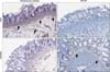 | Fig. 1Hirschsprung’s disease (HD) case shows diffuse proliferation of acetylcholinesterase (AchE) fibers (arrows) in lamina propria, muscularis mucosa, and submucosa (A), but complete absence of calretinin immunoreactive fibers (C). Non-HD case reveals many thin calretinin immunoreactive fibers (arrows; D), but no proliferation of AchE fibers (B). (A, B) AchE stain, ×100. (C, D) Calretinin stain, ×100.
|
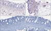 | Fig. 2Hirschsprung’s disease case shows equivocal acetylcholinesterase (AchE) findings which have mild proliferation of AchE fibers in submucosa (A, B; AchE stain, ×100), but complete absence of calretinin immunoreactive fibers was observed (C, D; calretinin stain, ×100). This case is difficult to differentiate diagnosis for normal variation.
|

Notes
References
1. Langer JC. Chapter 101 Hirschsprung disease. In : Stehr W, Gingalewski CA, editors. Pediatric surgery. 7th ed. Philadelphia: Elsevier Saunders;2012. p. 1265–1278.
2. Langer JC. Chapter 34 Hirschsprung disease. In : Holcomb GW, Patrick Murphy J, Ostlie DJ, editors. Ashcraft’s pediatric surgery. 6th ed. London, New York: Elsevier Saunders;2014. p. 474–491.
3. Stewart CL, Kulungowski AM, Tong S, Langer JC, Soden J, Sømme S. Rectal biopsies for Hirschsprung disease: patient characteristics by diagnosis and attending specialty. J Pediatr Surg. 2016; 51:573–576.

4. Langer JC. 50 Years ago in the Journal of Pediatrics: rectal biopsy as an aid in the diagnosis of diseases of infants and children. J Pediatr. 2013; 162:301.
5. Meier-Ruge W, Lutterbeck PM, Herzog B, Morger R, Moser R, Schärli A. Acetylcholinesterase activity in suction biopsies of the rectum in the diagnosis of Hirschsprung’s disease. J Pediatr Surg. 1972; 7:11–17.
6. Park WH, Choi SO, Kwon KY, Chang ES. Acetylcholinesterase histochemistry of rectal suction biopsies in the diagnosis of Hirschsprung’s disease. J Korean Med Sci. 1992; 7:353–359.
7. Lake BD, Malone MT, Risdon RA. The use of acetylcholinesterase (AChE) in the diagnosis of Hirschsprung’s disease and intestinal neuronal dysplasia. Pediatr Pathol. 1989; 9:351–354.
8. Barshack I, Fridman E, Goldberg I, Chowers Y, Kopolovic J. The loss of calretinin expression indicates aganglionosis in Hirschsprung’s disease. J Clin Pathol. 2004; 57:712–716.
9. Eusebi P. Diagnostic accuracy measures. Cerebrovasc Dis. 2013; 36:267–272.
10. Park WH. Clinical features and diagnosis of Hirschsprung’s disease. J Korean Assoc Pediatr Surg. 2002; 8:48–53.
11. Cinel L, Ceyran B, Güçlüer B. Calretinin immunohistochemistry for the diagnosis of Hirschprung disease in rectal biopsies. Pathol Res Pract. 2015; 211:50–54.
12. Kapur RP, Reed RC, Finn LS, Patterson K, Johanson J, Rutledge JC. Calretinin immunohistochemistry versus acetylcholinesterase histochemistry in the evaluation of suction rectal biopsies for Hirschsprung disease. Pediatr Dev Pathol. 2009; 12:6–15.
 XML Download