

 PDF
PDF ePub
ePub Citation
 Print
Print


INTRODUCTION
Actinomycosis is a relatively rare and chronic progressive suppurative disease that is mainly caused by Actinomyces israelii. A. israelii is an anaerobic Gram-positive organism that is normally present in the oral cavity as well as throughout the gastrointestinal tract, female genital tract, and the bronchus [1] that spreads by direct extension across the tissue planes with the formation of multiple abscesses, abundant granulation tissue, and draining sinuses [2]. Actinomycosis occurs most frequently in the cervical facial (50% to 65%), abdominal (20%), and thoracic (15%) regions [1].
Abdominal actinomycosis, the incidence of which has increased over the past few years [3], usually involves the vermiform appendix and terminal ileum [34]. Its clinical symptoms are non-specific and include abdominal pain, fever, nausea, vomiting, and diarrhea [3]. If the appendix ruptures, right-sided pain and other symptoms of appendicitis can develop [345]. A combination of antibiotics and surgical intervention is the treatment of choice for abdominal actinomycosis [6].
Here we describe an 18-year-old girl who underwent a laparoscopic exploration for acute appendicitis and was diagnosed with appendiceal actinomycosis upon histopathological examination.
CASE REPORT
An 18-year-old girl who had a 4-day history of right lower abdominal pain visited at a nearby hospital, and she had undergone laparoscopic exploration for a presumed diagnosis of acute appendicitis. At that hospital, an unexpected mass-like lesion beneath the cecum in the right lower quadrant (RLQ) was found, and she was transferred to our hospital for further evaluation.
Physical examination revealed tenderness in the RLQ and no rebound pain. There were no associated symptoms such as fever, loss of appetite, nausea or vomiting, or diarrhea. She had no relevant past or family history.
Laboratory findings included mild leukocytosis (11,760/µL; neutrophils 79.7%, eosinophils 0.8%) and an elevated C-reactive protein level (3.09 mg/dL).
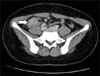
A contrast-enhanced abdominal CT scan revealed a 4.3×2.3 cm mass-like lesion beneath the cecum in the RLQ and an invisible normal appendix (Fig. 1).
From these findings, acute appendicitis with a peritoneal abscess was primarily diagnosed and a laparoscopic exploration was planned.
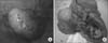
Operative findings showed a 3×3 cm appendiceal mass with dense severe adhesions (Fig. 2A, B) for which we performed an ileocecectomy.
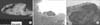
Gross pathology revealed severe inflammation and adhesion between the appendix and the adjacent appendiceal serosal surface. Irregular nodularity was seen in the distal portion of the ileal mucosal area, while intraluminal obliteration was found due to the severe inflammation (Fig. 3A).
Light microscopic examination revealed acute suppurative appendicitis with a periappendiceal abscess and actinomycotic colonies. At higher magnification, a typical sulfur granule surrounded by neutrophils was found that was confirmed as appendiceal actinomycosis (Fig. 3B, C).
After the operation, she was administered intravenous antibiotics (cefotaxime, metronidazole, piperacillin/tazobactam) for 20 days and then oral amoxicillin 1 g three times a day for at least 3 months.
DISCUSSION
Abdominal actinomycosis is a rare and insidious disease [78]. The overall incidence of actinomycosis is virtually impossible to ascertain since failure to consider it as a diagnosis and difficulty confirming it lead to under-reporting [9]. Approximately 65% of abdominal actinomycosis cases occur in association with acute appendicitis [10].
The symptoms, signs, and laboratory findings of abdominal actinomycosis are nonspecific, while its radiologic findings often show a poorly defined mass with irregular margins [11]. As such, its diagnosis is often delayed. Only 10% of cases are diagnosed preoperatively, and it is difficult to differentiate abdominal actinomycosis from a malignant tumor or appendicitis [7].
The differential diagnosis of abdominal actinomycosis should include Crohn's disease, ulcerative colitis, diverticulitis, neoplasms, and intestinal tuberculosis [811]. The definitive diagnosis of actinomycosis requires microscopic proof of either the pathogen itself or the presence of specific "sulfur granules" [8].
Tissue sampling and a histologic examination are important to ensuring a correct diagnosis, but the former is not always possible for two reasons. First, the clinical presentation of the disease may be acute or the sample may be unobtainable using percutaneous methods. Second, Actinomyces is a Gram-positive, slow-growing, non-acid-fast, anaerobic, and filamentous bacterium that requires specific incubation in special culture medium for at least 7 days [1]. Accordingly, culture results are negative in 76% of cases [12] and excision biopsy for histopathological examination is frequently needed [1].
The combined use of surgery and penicillin therapy has a reported cure rate of 88.8% [13]. Antibiotic therapy is useful for preventing the local recurrence of actinomycosis. Penicillin in particular has been shown to be effective in treating abdominal actinomycosis. Other commonly used antibiotics include tetracycline, erythromycin, doxycycline, clindamycin, imipenem, ceftriaxone, and ciprofloxacin [7].
A combination of long-term antibiotic therapy and adequate surgery is necessary to ensure the complete eradication of actinomycosis because of the large degree of reactive fibrosis formed by the infection [14]. However, recent studies have shown that a combination of complete surgical resection followed by short-term antibiotic treatment is effective [15].
Similarly, in this case, as the disease does not present with any typical features, either clinically or radiologically, the patient was initially diagnosed with appendicitis, and received laparoscopic exploration for a presumed diagnosis of acute appendicitis. The diagnosis was made after pathologic examination of the specimen, and then antibiotics were changed according to the pathologic results.
In summary, abdominal actinomycosis is difficult to diagnose preoperatively due to its atypical symptoms and imaging findings. However, since its accurate diagnosis is important to the prevention of unnecessary surgical treatment or to adequate antibiotic therapy, abdominal actinomycosis should be included as a differential diagnosis when an unusual abdominal mass or abscess presents on abdominal CT.
 XML Download