

 PDF
PDF ePub
ePub Citation
 Print
Print


INTRODUCTION
Remnants of vitellointestinal duct (VID) account for a wide variety of umbilical abnormalities. These include fistula, sinus tract, umbilical adenoma, enterocystoma and congenital bands [1]. Incidence of patent vitellointestinal duct (PVID) is reported as 0.0053% [2]. Furthermore, bowel prolapse through PVID is rare and double prolapse of proximal as well as distal loop in a newborn is extremely rare. Omphalocele with prolapsing bowel through PVID as found in our index case is even rarer.
CASE REPORT
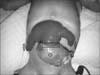
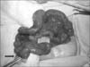
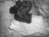
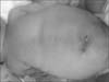
A full term male neonate, weighing 2,400 g, delivered by normal spontaneous vaginal delivery presented to our institute within 3 hours after birth; at first glance, he had a protruding mass through an umbilical defect covered with thin membrane and also had some prolapsed bowel loops. During the antenatal period, he was supervised with antenatal ultrasounds being normal. On examination, the cry, tone and activity of the baby were good. There was an omphalocele minor covered with a thin membrane and on the top of it were some prolapsed intestinal loops giving shape of an inverted Y (Fig. 1). Meconium could be seen coming from the proximal limb of this Y. Patient was taken up for surgery after adequate hydration. Under general anesthesia, prolapsed loops were approached through circumferential incision and excision of omphalocele membrane. Intestine was delivered outside the abdomen and prolapsed bowel was reduced. A PVID opening was noted in the distal ileum at the site of prolapse (Fig. 2). It was a wide mouthed PVID from where the ileal loops were intussuscepting and prolapsing outside. The intussuscepting bowel loop was gently reduced. The distal ileal segment with the PVID was resected and an end to end anastomosis of the bowel was done (Fig. 3). The omphalocele had only distal part of ileum, entire cecum and ascending colon as its contents. These contents were replaced into the abdomen and umbilicoplasty was done with absorbable sutures (Fig. 4). Postoperative period was uneventful. Patient was started on small amount of feeds on 5th postoperative day, and discharged on full breast feeds on 13th postoperative day. At 3 months follow-up, the child is thriving well.
DISCUSSION
The VID connects the yolk sac to the gut in a developing embryo and provides nutrition until the placenta is established. Between the 5th and 7th week of gestation, the duct attenuates and separates from the intestine. Just before this involution, the epithelium of the yolk sac develops a lining similar to that of the stomach. Partial or complete failure of involution of the VID results in various residual structures. Meckel's diverticulum is the most common of these structures and is the most frequent congenital gastrointestinal anomaly occurring in 2% to 3% of all infants. Other VID remnants occur infrequently, including a persistently patent duct, a solid cord, or a cord with a central cyst or a diverticulum associated with a persistent cord between the diverticulum and the umbilicus [34]. Bowel prolapse through PVID is rare and double prolapse of proximal as well as distal loop in a newborn is extremely rare [56]. Furthermore, omphalocele with prolapsing bowel through PVID as seen in our index case is even rarer. After a thorough search of literature, we came across only five similar case reports [7891011].
The condition, if not managed promptly by surgical intervention, may lead to subacute or acute intestinal obstruction, strangulation and gangrene of the prolapsed intestinal loops. Primary closure of the PVID following reduction of the prolapsed intestines may be possible if the patient arrives early without any gross edema over the intestinal loops. If the defect is large one can go for resection of the loop of intestine near the patent duct followed by primary anastomosis. If the patient arrives late with gross edema or the viability of the intestinal loops is in question then exteriorization of the suspected loop or loop ileostomy is advised [12]. The good result with early primary resection and anastomosis in our case emphasizes the importance of prompt referral, early diagnosis and quick surgical intervention to prevent edema and gangrene which can be fatal. Even the morbidity associated with a stoma can be prevented if the pediatric surgeon is familiar with this unique presentation and decides to intervene early based on his clinical examination.
PVID with prolapsed ileal loop is a rare condition which needs prompt diagnosis, surgical reduction and repair of the defect. Since other malformations are not present, this entity may be regarded as a distinct entity, a form of a lower midline abdominal wall defect understood by a disturbance of development of vitelline duct and having excellent prognosis.
 XML Download