

 PDF
PDF ePub
ePub Citation
 Print
Print


Abstract
Palpable inguinal mass in children should be differentiated from inguinal hernia, hydrocele, lymph node, and tumor. Though using ultrasonography, fatty tumor would be misdiagnosed as incarcerated inguinal hernia containing fatty component. We experienced the huge inguinal lipoblastoma in 5-year-old girl mimicking recurrent incarcerated hernia. Laparoscopic exploration revealed it was not incarcerated hernia but well demarcated bulging mass from abdominal wall. Mass was about 10×4×3 cm and extended from internal inguinal ring to saphenous opening. It was near total excised because of right external iliac vein injury. Pathologically, it was proven as lipoblastoma containing mature adipocyte with lipoblast and fibrous septa. Postoperatively, we noticed a segmental thrombotic occlusion of external iliac vein. After 1 year, she has no symptom related to occluded vessel. The remained lipoblastoma showed no interval change. Even lipoblastoma has a good prognosis with low recurrence rate, we need careful follow-up.
Figures and Tables
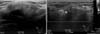 | Fig. 1Ultrasonography showed abnormal echogenic fatty mass in the right inguinal fossa (3×1.3×4.3 cm) with minimal movement during Valsalvar's maneuver.
|

Notes
References
1. Kamal NM, Jouini R, Yahya S, Haiba M. Benign intrascrotal lipoblastoma in a 4-month-old infant: a case report and review of literature. J Pediatr Surg. 2011; 46:E9–E12.
2. Chung EB, Enzinger FM. Benign lipoblastomatosis. An analysis of 35 cases. Cancer. 1973; 32:482–492.
3. Coffin CM. Lipoblastoma: an embryonal tumor of soft tissue related to organogenesis. Semin Diagn Pathol. 1994; 11:98–103.
4. Burchhardt D, Fallon SC, Lopez ME, Kim ES, Hicks J, Brandt ML. Retroperitoneal lipoblastoma: a discussion of current management. J Pediatr Surg. 2012; 47:e51–e54.
5. Nam SH, Kim DY, Kim SC, Kim IK. The clinical manifestations of lipoblastoma in children. J Korean Assoc Pediatr Surg. 2007; 13:179–186.
6. Hicks J, Dilley A, Patel D, Barrish J, Zhu SH, Brandt M. Lipoblastoma and lipoblastomatosis in infancy and childhood: histopathologic, ultrastructural, and cytogenetic features. Ultrastruct Pathol. 2001; 25:321–333.
7. Pham NS, Poirier B, Fuller SC, Dublin AB, Tollefson TT. Pediatric lipoblastoma in the head and neck: a systematic review of 48 reported cases. Int J Pediatr Otorhinolaryngol. 2010; 74:723–728.
8. Dilley AV, Patel DL, Hicks MJ, Brandt ML. Lipoblastoma: pathophysiology and surgical management. J Pediatr Surg. 2001; 36:229–231.
9. Mognato G, Cecchetto G, Carli M, Talenti E, d'Amore ES, Pederzini F, et al. Is surgical treatment of lipoblastoma always necessary? J Pediatr Surg. 2000; 35:1511–1513.
10. Lorenzen JC, Godballe C, Kerndrup GB. Lipoblastoma of the neck: a rare cause of respiratory problems in children. Auris Nasus Larynx. 2005; 32:169–173.
 XML Download