

 PDF
PDF ePub
ePub Citation
 Print
Print


INTRODUCTION
Gastroschisis and omphalocele are two common congenital anterior abdominal wall defects. Omphalocele is a failure of the intestine to return to the abdominal cavity through the umbilicus, occurring at approximately 12 weeks of gestation [1]. The defect involves the umbilicus, and the herniated viscera are covered with a membranous sac. Gastroschisis is a frank defect in the abdominal wall through which the viscera prolapses usually to the right of the umbilical cord. Because the peritoneal sac is absent, the fetal bowel is continuously exposed to the amniotic fluid, resulting in significant inflammation of the bowel wall [2]. Omphalocele is known to have more associated anomalies and higher mortality rate than gastroschisis [2]. The purpose of this study is to analyze the clinical differences between gastroschisis and omphalocele with a special emphasis on outcome.
METHODS
The study group consisted of 103 live infants with congenital abdominal wall defects detected at delivery in Asan Medical Center between September 1989 and February 2013. Data were retrospectively obtained and reviewed from the medical records.
In all cases of gastroschisis, we tried to do early primary repair because there was no membrane covering of the exposed organs. However, if abdominal pressure was expected to increase to impede respiratory and circulatory function with primary repair, the abdominal wall was covered temporarily with artificial material. The same practice was applied to omphalocele, but the 1st operation was delayed in some patients who had large defect and showed unstable vitality. The operative strategy was based on the clinical decision of the surgeon according to the condition of a patient, irrespective of the defect size.
All statistical analyses were performed using PASW Statistics version 18.0 (IBM Co., Armonk, NY, USA). The chi-square or Student's t test was done. Statistical significance was accepted for p<0.05.
RESULTS
Over the 24-year period, 103 cases of congenital anterior abdominal wall defects were encountered with gastroschisis in 43 cases and omhalocele in 60 cases. The prenatal diagnosis was identified in 27 cases of gastroschisis and 32 cases of omphalocele.
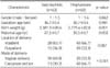
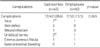
There was a female predominance within both groups, but this did not reach statistical significance. Gestation ages showed no difference. The mean birth weights were 2,381.9±80.6 g for gastroschisis and 2,779.4±82.8 g for omphalocele (p=0.001). Mean maternal ages in the gastroschisis and omphalocele groups were 27.5±0.7 years and 30.5±0.7 years, respectively (p=0.002). The preferred mode of delivery was vaginal delivery in gastroschisis, but Caesarean section (C/S) in omphalocele (Table 1).
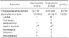
Associated malformations were documented in 46 of the 60 infants with omphalocele, while in 13 of the 43 infants with gastroschisis (Table 2). Cardiac anomalies were most frequently associated in both groups. Gastrointestianl abnormalities were ileal atresia (1) and imperforate anus (1) in gastroschisis, and ileal atresia (1) and imperforate anus (3) in ompahlocele, excluding intestinal malrotation. There were chromosomal abnormalities in 8 among 20 omphalocele cases, including Beckwith-Wiedemann syndrome (5), 13 trisomy (1), 47 XYY (1), and uniparental disomy 14 (1). Chromosomal analysis was done in seven cases of gastroschisis, which showed no abnormality.
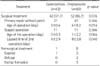
Surgery was done in 42 cases (97.7%) of gastroschisis and 52 cases (86.7%) of omphalocele. One patient with gastroschisis died before surgery due to respiratory failure. In 8 cases of omphalocele, surgery was not performed due to early neonatal death (3), parental refusal (2), or eschar formation (3). One-stage primary repair without prosthesis of the abdominal wall defect was achieved in 31 cases of gastroschisis. Staged procedures were carried out in 11 cases, and the mean interval between operations was 4 days. In 41 cases of omphalocele, one-stage primary repair without prosthesis of the defect was possible at the age of 0 to 40 days. In 11 cases, staged surgical procedures were carried out and the mean interval between operations was 8 days. The abdominal wall defect size varied between 2 and 15 cm. In case of staged operation, abdominal wall defect was temporarily closed with saline bag sheet, polytetrafluoroethylene (PTFE) patch or adhesive surgical drape at the first operation. Primary repair was done earlier in gastroschisis (p<0.05). The lapsed time from 1st to 2nd operation in staged repair was longer in omphalocele (p<0.05) (Table 3).
The mortality rate of gastroschisis and omphalocele were 23.3% (10/43 cases) and 15.0% (9/60 cases), respectively (p=0.287). The main causes of death were abdominal compartment syndrome (6/10 cases) in gastroschisis, and respiratory failure (4/9 cases) and discharge against medical advice (4/9 cases) in omphalocele (Table 4).
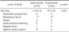
In survived patients, postoperative complications occurred in 12 patients with gastroschisis and 7 patients with omphalocele (Table 5). In the gastroschisis group, ileus was the most common postoperative complication. There was no difference between the primary repair group and the staged operation group. Two ileus cases required surgical treatment. The wound infection was complicated in 3 cases of omphalocele with one of primary repair and two of staged operation.
DISCUSSION
The prevalence of omphalocele has been reported to be 1.22-2.76 per 10,000 births, and gastroschisis, 0.94-3.01 per 10,000 births [3,4,5]. The exact etiology of these anomalies is unknown. Unlike gastroschisis, the prolapsed organs in omphalocele are covered with a protective sac [6,7]. Maternal age tends to vary at the extremes of the spectrum in omphalocele with the risk of very young and more advanced maternal age. Gastroschisis is associated with very young maternal age in the literature (22-23.6 years) [8,9]. In this study, the maternal age of gastroschisis was younger than that of omphalocele group.
The preferred mode of delivery is controversial. Routine C/S was not justified [10]. It was noted that there was no benefit from C/S for gastroschisis; instead, the high rate of respiratory distress, gastrointestinal dysfunction, and bowel stenosis were observed when C/S operations were performed only for obstetric reasons. In our patients, 30% of the infants with gastroschisis and 67% of the infants with omphalocele were delivered by C/S, which meant C/S was the main mode of delivery in omphalocele for the fear of sac rupture.
Omphalocele is frequently seen in the context of congenital syndromes with multiple associated anomalies, such as Beckwith-Wiedemann syndrome, cloacal exstrophy, OEIS (omphalocele, exstrophy, imperforate anus, spinal defects) complex, Cantrell's pentalogy [3,4]. Cardiac anomaly is the most frequent anomaly in omphalocele (39%-45%) as in our study [11,12]. Omphalocele is often associated with chromosomal abnormalities, such as trisomies 13 and 18 [13,14]. In our series, there were no karyotypic abnormalities in 7 cases of gastroschisis, but there were karyotypic abnormalities in 8 among 20 omphalocele patients.
In gastroschisis and omphalocele, the herniated viscera should be reduced into the abdomen and a solid abdominal wall be created. The surgery should be performed in a manner to minimize risks of increased abdominal pressure to the baby. However, if abdominal pressure is expected to increase to impede respiratory and circulatory function with primary repair, the abdominal wall can be covered temporarily with artificial material. This silo covers the extra abdominal organs and protects them from infection or loss of fluids. After a few days, the organs decongest, and the abdominal wall repair can then be tried. For silo formation, we used adhesive surgical drape, PTFE or normal saline bag sheet. Eschar formation with preservation of omphalocele sac and delayed repair is a good option in a case of severe unstability or huge omphalocele.
The mortality of these conditions has decreased markedly in recent years [12]. This is attributable to improved prenatal diagnosis, advances in surgical technique and pre- and postoperative cares [15]. Prenatal diagnosis made it possible to manage mother and neonate in one perinatal center. Therefore, the delivery and subsequent surgery can be planned and carried out in an interdisciplinary fashion. The mortality rate of omphalocele has been well known to be higher than that of gastroschisis [5,9,15,16]. Many studies demonstrated that this higher mortality was attributed to higher association of cardiac anomaly in omphalocele [17,18]. In our result, the mortality rate of omphalocele was lower than that of gastroschisis (15.0% vs. 23.3%), even though it was not significant. The explanation of this lower mortality rate might be the improvement of treatment of cardiac anomalies, because no patient died from cardiac dysfunction in our study. Furthermore, the compartment syndrome as the main cause of death in gastroschisis should be considered for the comparison of the mortalities.
 XML Download