

 PDF
PDF ePub
ePub Citation
 Print
Print


Introduction
Quadriceps femoris is the powerful extensor muscle of knee joint present in the anterior compartment of thigh. It usually arises by four heads namely rectus femoris (RF) from anterior inferior iliac spine and from a groove above the acetabulum where as vastus lateralis (VL), vastus medialis (VM), and vastus intermedius (VI) arises from the upper two-third of the femoral shaft apart from the linea aspera and cover it from trochanter to condyles [1]. Distally these four tendons unite to form a single tendon to get inserted into base of patella. But, recent cadaveric study by Grob et al. [1] in Swiss population has discovered an additional new muscle namely tensor of vastus intermedius (TVI) which is situated between VL and VI. The TVI muscle arises from the anteroinferior part of greater trochanter and is inserted in the upper part of patella. The presence of the additional muscle can alter the mechanics of the patella and also have an impact on the surgeries of extensor apparatus of knee. The description and knowledge regarding the attachments, variability and morphometry of TVI in Indian population is lacking in the literature. Hence, this study was taken up to investigate the TVI muscle in South Indian population and to describe its variation.
Materials and Methods
The data for the present study was collected from formalin fixed adult cadavers allotted for undergraduate dissection in the Department of Anatomy, retrieved from the institutional body donation program following the ethical guidelines. A total of 36 lowerlimbs were dissected. This included 27 male lowerlimbs (8 paired and 11 unpaired) and nine female lowerlimbs (3 paired and 3 unpaired). A longitudinal incision was made on the front of thigh from midinguinal point to patella. Two transverse incisions made one at the inguinal region and the apex of patella. The skin was reflected and the superficial fascia was removed. The deep fascia was cleared. The quadriceps muscle was identified and the components of the muscle were defined by blunt dissection. The sartorius and RF muscle were transected distally and reflected. The TVI muscle was identified between the VL and VI in the proximal part. The anatomy of the TVI was studied with respect to its location, origin, point of fusion of its aponeurosis and the neurovascular pedicle to the muscle. The TVI was classified according to the classification by Grob et al. [1]. The following measurements were taken namely length of the muscle belly (distance between most proximal and most distal points at which muscle fiber can be seen), length of the aponeurosis (most distal point of muscle fiber to the superior aspect of patella) and the distance of fusion of TVI from superior aspect of patella to VL or VI were measured with Mitutoyo digital vernier caliper (Mitutoyo, Tokyo, Japan) to the nearest millimeter. The measurements were made by a single investigator thrice and the mean of it was taken as the final measurement.
Cadavers with any lower limb anomalies, hematomas, fractures, tumors, or lacerations were excluded from the study. All the dissected specimens were photographed with a digital camera. The results were statistically analyzed for the following parameters namely mean, standard deviation, and significance of side.
Results
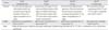
A total of 36 lower limbs have been dissected (27 male and 9 female). In all the dissected lower limbs, the TVI muscle was identified. The identified muscle was located between VI and VL (Fig. 1). The morphology of the muscle is classified into four types mainly based on the aponeurosis course [1]. However, in the present study, the origin of the TVI and its aponeurosis course is taken into consideration for the classification as as follows.
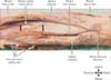
Type 1 (independent): the muscle takes origin from the upper part intertrochantric line and anterior part of greater trochanter but the origin is separable from VL origin (33.33%). The aponeurosis is separable from both VI and VL (Fig. 1).
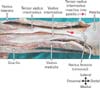
Type 2 (VI type): the muscle takes origin together with VI and posterior border of TVI is fused with VI (8.33%). The aponeurosis is separable from VL (Fig. 2).
Type 3 (VL type): the muscle takes origin from the VL and the origin is inseparable (30.56%). The aponeurosis is separable from VI (Fig. 3).
Type 4 (common): the muscle takes origin from the VL and the origin is inseparable (27.78%). The aponeurosis is separable both VL and VI (Fig. 4).
In three of the dissected cadavers, the TVI muscle arises by two heads.
Distal course
Distally the aponeurosis is attached to the upper medial aspect of patella fusing with either the VL, VI, RF, or a combination of two or all the three aponeurosis (Fig. 5). The course of the TVI distally is such that the ventral border becomes medial and the dorsal border becomes lateral. The fusion of the lateral border of TVI was with VL or VI was 26%. The fusion of TVI with VL and VI or VL and RF or VI and RF was 66.67%. The fusion of TVI to VL, VI, and RF was 8.33%. The branches of femoral nerve and vessels were identified separating the TVI muscle belly from VL (Fig. 1).
Quantitative data
The mean and standard deviation of the length of TVI was 145.40±37.55 mm. The mean and standard deviation of length of TVI of right and left side were 142.91±39.37 mm and 148.23±36.40 mm, respectively. The sample included only nine female lower limbs but however the length of the muscle was more in female cadaveric lowerlimbs (162.59±47.41 mm) compared to males (139.70±32.72 mm).
The mean and standard deviation of the length of the aponeurosis of TVI was 193.55±42.32 mm. The mean and standard deviation of length of the aponeurosis of TVI muscle of right and left side were 196.41±45.05 mm and 190.35±40.18 mm, respectively. Similar to the length of the muscle, the length of aponeurosis was also more in females (200.87±35.64 mm) compared to males (191.11±44.68 mm).
The distance of fusion of TVI from superior aspect of patella to VL or VI was 98.42 mm with a wide range (maximum value was 170.84 mm and minimum value was 23.19 mm).
There was no significant difference in the length of the TVI muscle and its aponeurosis length between the right and the left sides.
Discussion
Willan et al. [2] was the first to report an additional muscle lamina between the tendon of VL and VI. However the further description of a new muscle between VL and VI, referred as TVI was documented by Grob et al. [1]. The new muscle TVI was classified into four types by Grob et al. [1] namely independent type, VL type, VI type, and common type. It was documented that the origin of the muscle was from anterior aspect of greater trochanter and distal to intertrochantric line [1]. However, the origin of the TVI in the individual types have not been described in detail. In the present study, the origin of the muscle, its middle and distal course of the individual types has been documented in detail (Table 1). The origin of the muscle was from the upper part of intertrochanteric line and greater trochanter of femur only in independent type of TVI (33.33%). In all other types, the origin of TVI was either together with VL or VI which was 66.67% in the present study. The middle third of the muscle in the present study was similar to the findings of the previous study [1]. The distal course of the TVI inserted into the upper medial aspect of patella fusing with either the VL, VI , RF, or a combination of two or all the three aponeurosis. In the present study, the fusion of lateral border of TVI was with VL and VI or VL and RF or VI and RF (66.67%). In most of cadavers, the fusion of the lateral border of TVI was with VL and RF.
The quantitative data of the TVI was not available in literature for comparison. However, the point fusion of TVI from superior aspect of patella to VL or VI is significant as the tensor action of the small muscle depends probably on the distance of fusion. The tensor action of the small muscle TVI can be significant if the distance of fusion of the aponeurosis is close to patella. As VL causes the patella to move laterally and VM especially vastus medialis oblique (VMO), pulls the patella medially both just aligns the patella within the trochlear groove as the knee moves through flexion and extension. Though VMO is considered the primary medial stabilizer of the patella, it has little function in knee extension [3456]. The morphology of TVI, its course from lateral to medial aspect of thigh to get inserted into the medial aspect of upper border of patella overlying on VI can impose considerable tension on VI to medialise its action. The fusion of TVI aponeurosis distally with the adjacent muscles VL, VI, and RF may play a significant role in knee extension. Furthermore, it is documented that the importance of TVI in a patient with knee pain associated with rupture of the TVI tendon and was supported by magnetic resonance imaging evidence [7]. Sonographic identification of the muscle has been documented and its concluded that the TVI should not be mistaken for focal tendinitis due to course of the muscle from lateral to medial and giving the appearance of focal thickening of quadriceps tendon [8]. The cellular precursor of the limb muscles are located in the ventrolateral half of somite which migrate into limb buds. During a certain stage of development, the muscle primordia within the different layers of the thigh fuse to form a single muscle mass; thereafter, some muscle primordia disappear through apoptosis. Though there is no definitive literature regarding the embryology of different types of TVI, it can be attributed to the degree of disappearance of muscle primordial on the lateral aspect of thigh. However, further research is needed to establish the embryological and genetic basis of the different types of TVI.
The TVI muscle which is a part of the quadriceps apparatus could play a significant role in knee extension and alignment of patella which can be crucial in isolated traumatic injury to its aponeurosis or the muscle. The exact role of TVI on the patella and its role in knee extension need to be investigated further.
The TVI muscle was identified in all the dissected cadavers of this population and the most common type being “independent type” of TVI with neurovascular pedicle from lateral circumflex branch of femoral artery and posterior division of femoral nerve. The muscle origin was from the upper part of intertrochanteric line and anterior part of greater trochanter of femur inserted to medial aspect of upper border of patella. The variations in types and the quantitative data can add to the existing knowledge and serve as anatomical basis to understand its function and mechanics on the patella.

 XML Download