

 PDF
PDF ePub
ePub Citation
 Print
Print


Introduction
The dysfunction of reproductive system and infertility are the most important adverse effects of chemotherapy [1]. Busulfan (1,4-butanediol dimethanesulfonate) is a bifunctional alkylating agent, that use to treat chronic myeloid leukemia and before a bone marrow or stem cell transplant, which inhibits cell division by sticking to one of the DNA strands. Therefore, the tissues such as bladder, liver, skin, nervous system, and gonadal and cells with high division activities like as testes and germ cells and somatic cells are influenced by adverse effects of busulfan. In addition, treatment by busulfan affects fertility in the male and makes prolonged and sometimes irreversible depression of sperm counts in humans [23]. It has been show that the single doses of busulfan can preferentially kills spermatogonial stem cell and cause sterilize mice by deplete germ cells and disrupt the junctions between Sertoli cells, and cause long-term morphological damage to sperm produced by surviving spermatogonia [1]. Effects of busulfan on the spermatogenesis have been evaluated on testis and male reproductive system [345], but the effects of busulfan on sperm structure are still unclear. However, more information about the effect of chemotherapeutic agent on sperm morphology and structure is needed to treat these infertile men [5678].
Abnormal morphology of spermatozoa can be related to the male infertility. In the last years, there has been much debate on a new potential parameter and methods of sperm morphology analysis and relation of these parameters by male fertility. The human sperms dimension and their potential roles in male infertility are not still fully understood [1]. Therefore, evaluation of sperm morphology can be considered as one of the most important factors for a successful fertilization and determining sperm quality [9101112]. Although the World Health Organization (WHO) recommendations to evaluation various other constituent part of sperm morphology but little attention has been paid to the parameters of the volume of sperm's head and length of sperm's mid-piece and sperm's tail, in spite of they play a part in to sperm motility [1314]. Thus, studies can have been constrained to special areas; including sperm tail and sperm head [1516]. Therefore, measurement of the volume of sperm's head and length of sperm's mid-piece and sperm's flagellum can be useful for to known sperm quality. Stereological methods considered in this study because unbiased stereological techniques were used in this study are easy, fast and accurate [11]. In the light of the above-mentioned reports, this investigation was designed to evaluate the effects of busulfan on the volume of sperm's head and length of the sperm's tail and mid-piece by using stereological methods.
Materials and Methods
In this study, 12 adult mice (28–30 g) were obtained from the laboratory animal center of Shahid Beheshti University of Medical Sciences, Tehran, Iran. The Ethics Committee of the University approved the animal experiment (IR.SBMU.SM.REC.1395.584). The mice were assigned to tow experimental groups: (1) control and (2) busulfan. Each group included six mice that were housed under standard conditions, room temperature (22℃–24℃), and a 12:12 hour light-dark schedule and had free access to water and food.
Busulfan treatment
Oligospermia can be induced in mice by single dose (20 mg/kg) intraperitoneal injection of the busulfan. All mice were kept for 8 weeks after the busulfan injection then sperm morphology examination was done.
Stereological study
Estimation of mean volume of sperm's head
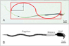
The volume was estimated using the nucleator method. The sperm's head was cut into isotropic uniform random pieces using the orientator method. They were stained with Diff-Quik. The sperm's head were sampled using an optical dissector. For each sampled sperm's head, two horizontal directions (intercept, ln) were considered from the central point within the sperm's head to the sperm's head borders. From a series of these measurements (120–200 intercepts in each group), the mean sperm's head volume in the number weighted distribution was estimated [1819] (Fig. 2):

Estimation of the length of sperm's mid-piece and flagellum
Each slide was studied with a video-microscopy system made up of a microscope (E-200, Nikon, Tokyo, Japan) linked to a video camera. An oil immersion objective lens ×100 was used to achieve a better recognition of the head, mid-piece and flagella. The method of estimation of the length of sperm's mid-piece and flagella on microscopic slides is an example of the application of length estimation in 2D space. The whole slide was covered by sampling it in a systematic random manner. Briefly, the microscopic fields were sampled in a systematic random manner, by moving the microscope stage in an equal interval along the X- and Y-direction of the stage, using a stage micrometer. One hundred sperms were sampled on each slide (each subject). There are some guidelines that will suffice for stereological sample size to have an acceptable precision. According to these rules 100–200 probe interaction (e.g., sampling of 100 intersections of Merz grid with sperm's mid-piece and flagella) is sufficient. This procedure was continued until the whole slide was studied. In each sample field a test grid composed of two elements was superposed on the image on the monitor. If a sperm's head lay inside the frame and did not touch the forbidden lines (left and inferior borders of the frame), it was sampled. The mean sperm's mid-piece and flagella length was estimated using the following formulae [1819]:
where a/l is the Merz grid constant which was obtained as follows: the area of each basic tile of the grid was X multiplied by Y. Within this tile, there were two semicircles of length of π.d (perimeter of a circle), where d was the diameter of the semicircle. Thus the Merz grid constant a/l was (X_Y) π.d. asf was the area of the basic tile divided by the area of the counting frame. ΣI was the summation of the intersection of the tails with the semicircles. ΣN was the total number of the counted sperms in the unbiased counting frame (Fig. 2).
Results
Volume of the sperm heads
The results showed that the sperm heads volume was significantly different between control and busulfan groups (P<0.02) (Table 1). Therefore, sperm heads volume was associated with abnormal sperm morphology.
Discussion
The present study investigated the structural changes of the spermatozoa in the busulfan-induced oligospermia in rats using stereological methods. Our results showed a significant different in the volume and surface of the sperm's head in the control and busulfan groups. Our results also demonstrated that a significant different in the length of the sperm's flagellum in the control and busulfan groups. These changes in the sperm structural have been shown to be accompanied and related by impairments in the chance of sperm fertility [1]. Busulfan is a chemotherapeutic agent which has many side effects on reproductive system [2]. Busulfan is a potent agent, which causes the reduction and apoptosis of spermatogonial stem cells populations [2]. In the previous study showed that the morphological change of spermatozoa following administration of busulfan in rat [2]. Eight weeks after the intraperitoneal injection of busulfan, we found the structural changes in rat's spermatozoa. However, busulfan causes change and damage to the sperm structure when administered by an intraperitoneal injection [20]. Our results were in accordance with the previous studies reporting the adverse effects of busulfan [220] and showed that busulfan injections in male rat could induce azoospermia and structural changes in spermatozoa.
Moreover, there are several approaches for malignancy therapy such as chemotherapy and radiotherapy. It has been shown that fractionated irradiation results in depletion of endogenous spermatogenesis similar to using busulfan injection. There is little information about the mechanism of busulfan toxicity on the sperm structure, but its toxic effect on the reproductive system may associate with the fertility outcomes. Our result showed the relationship between busulfan toxicity and structural disorders in spermatozoa.
Busulfan can effect on the sperm structure such as volume of the sperm's head and the length of the sperm's flagellum. The reason why spermatozoa had abnormality in volume of the sperm's head and the length of the sperm's flagellum is unknown and need to more evaluation the mechanism of effect of busulfan on sperm structure. However, stereological methods can provide valuable quantitative information about the morphological changes in the sperm structure that cannot be obtained using sperm analysis by a time consuming other techniques like as image analyzers or tracing method on the electron or light microscopic samples. The stereological procedure not only faster than conventional methods but also no need special software to create these grids.
In conclusion, the use of stereological methods is recommended for the morphological change evaluation of the sperm by light microscopy. This method can to be a useful and efficient way to estimate the volume of sperm's head and sperm's mid-piece and tail lengths in rats.

 XML Download