

 PDF
PDF ePub
ePub Citation
 Print
Print


Introduction
Cecum is the blind pouch in the initial segment of large intestine inferior to ileocolic junction. Cecum and appendix are usually located in the right lower quadrant of abdomen [1]. Development of mid gut is a complex process which normally includes physiological herniation, rotation, retraction of loops, descent of cecum and fixation. Any abnormality in these processes may lead to malrotated mid gut or sub hepatic cecum [23]. Fundamental understanding of development of gastrointestinal tract helps in early diagnosis of malrotation of gut [1]. In case of appendicitis, with subhepatic cecum and appendix the diagnosis and surgical procedures are challenging. Malrotated mid gut may accompany with variation in blood supply, so surgeons should be aware of these to avoid undue complication during colonoscopy and surgeries. In the present case, we describe a subhepatic undescended cecum with its blood supply which is unique from previously reported cases.
Case Report
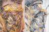
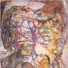
During the routine first-year undergraduate MBBS (Bachelor of Medicine Bachelor of Surgery) dissection, we found a rare developmental anomaly of undescended cecum in a male cadaver aged 60 years while demonstrating peritoneal folds. Further dissection was done to demonstrate its detailed anatomy and blood supply; then blood vessels were fabricated and photographed. In this cadaver, “conical” cecum was positioned in sub hepatic region measuring 4×3.5 cm (Fig. 1A). Appendix arising from the tip of cecum was located in 12'o clock position measuring 11.5 cm with “U” shaped bend at its tip (Fig. 1B). Ascending colon was very short measuring 4.5 cm in length and completely intraperitoneal. The 20-cm long terminal part of ileum extended from right lower quadrant to right upper quadrant to form ileocolic junction. External diameter of ascending part of terminal ileum is slender, measuring less than 1 cm, compared to the remaining part of terminal ileum having diameter of ~2 cm. Root of mesentery extended from duodenojejunal flexure up to terminal ileum in right lower quadrant normally and expanded toward subhepatic region by covering the slender terminal ileum up to ileocolic junction. Root of mesentery also extended up to cecum and ascending colon attaching it with posterior abdominal wall (Fig. 1B). The rest of hepatic flexure, colon and small intestine were normal. No fibrous Ladd's band was found between cecum and liver [4]. Blood supply to cecum and appendix was derived from ileo-colic artery, branch of superior mesenteric artery directed towards right upper quadrant. At the region, accessory right colic artery was found arising from ileo-colic artery (Fig. 2).
Discussion
At fifth to 10th week of intrauterine life, mid gut loops develop rapidly. As kidney and liver are simultaneously developing in the abdominal cavity with no space to accommodate all bowel loops, there is physiological herniation of mid gut loops. At tenth week, herniated loops are retracted in to abdominal cavity with total of 270° anti-clock wise rotation along the axis of superior mesenteric artery [5]. Reason for retraction is not well understood, it may be due to regression of mesonephric duct, decrease in liver size and growth of abdominal cavity. First part to re-enter the abdominal cavity is the proximal jejunum and last to enter is the cecal bud, so it arranges itself with jejunum to the left and cecum to the right of abdominal cavity. After retraction, cecum and appendix are placed in subhepatic region. At 11th week, cecum descends to the right iliac fossa by placing the hepatic flexure and ascending colon in right side of abdomen as in adult position [23]. Cecal bud from postarterial segment of midgut shows differential growth due to increase in diameter proximally and length distally resulting in development of distended cecal sac and narrow long slender appendix at birth. Then further differential growth in walls of cecum lead to the placement of opening of appendix posteromedially in cecum [23]. As cecum is completely intraperitoneal and devoid of mesentery its position in right lower quadrant is variable [6].
In the present case, midgut underwent 270° anti-clock wise rotation and cecum was fixed in subhepatic region. The causes of undescended subhepatic cecum reported in previous cases include Ladd's band, which is the fibrous band attaching from liver to cecum or ascending colon by obstructing the duodenum [7]. In 2013, Nayak et al. [8] reported subhepatic cecum with retroperitoneal terminal ileum as a cause of failure to descent. But in our case, there is no Ladd's band and the terminal portion of ileum is also completely intraperitoneal having mesenteric attachment up to ileocolic junction. The cause for undescended cecum in the present case may be due to the expansion of mesentery up to cecum leading to fixation and arrest of cecal descend at tenth week of intrauterine life. As cecum remains in conical form and ascending part of terminal ileum is slender, it's evident that the development of cecum and terminal ileum was arrested in intrauterine period due to persistence of mesenteric attachment. To the best of our knowledge, there have been no previous reported cases of undescended cecum with mesenteric fixation of cecum and terminal ileum.
A case of subhepatic cecum with appendicitis will not present with classical signs and symptoms; leading to delayed diagnosis of appendicitis or misdiagnosis as cholecystitis which may lead to perforation of appendix and surgical emergencies [9]. Appendicitis may also increase the chance of spread of infection to kidney, suprarenal gland, liver, and pancreas because of its subhepatic position.
During our literature search, we found a total of ten cases of subhepatic cecum reported from the year 1885 by Treves till now, but resources regarding blood supply of these subhepatic cecum and appendix are lacking, even though it has a lot of clinical significance during abdominal surgeries [10]. In one of the cases of subhepatic cecum, ileocolic artery was absent and cecum was supplied by right branches of middle colic artery and the tip of appendix was supplied by branch from right colic artery [10]. In the present case, superior mesenteric artery gives ileocolic trunk which in turns gives ileal branches to terminal ileum, ceacal branch, appendicular artery, and accessory right colic artery. Accessory right colic artery was supplying the very small ascending colon and hepatic flexure. The middle colic artery from superior mesenteric artery was supplying the transverse colon without giving right colic branch. Presence of accessory right colic artery is well documented in 10% of normal individuals, but till now no case of subhepatic cecum was reported with accessory right colic artery [1]. Variation in branching pattern of superior mesenteric artery in subhepatic cecum has great significance during emergency surgeries.
To summaries, subhepatic location of undescended cecum and appendix are rare developmental anomalies of mid gut. Surgeons and radiologists should be aware of these variations while interpreting clinical features and investigations to avoid delayed diagnosis or misdiagnosis. Knowledge about these arterial variations of such developmental anomalies is necessary to prevent undue complications and iatrogenic injuries during colonoscopy and surgeries.
 XML Download