

 PDF
PDF ePub
ePub Citation
 Print
Print


Introduction
Retinacula are thickenings of deep fascia in the region of joints, to keep the tendons in place as they cross the joints. Many retinacula are present in the vicinity of ankle joint. These retinacula are transverse bands across the ankle that holds down the tendons that cross from the leg to the foot. When we contract our leg muscles to move our foot or toes, the pull of the muscles on their tendons would raise these tendons away from the ankle, a phenomenon known as bowstringing which would weaken the ability of the muscles to move the foot or toes [1]. Superior and inferior extensor retinacula have been exhaustively described in the standard textbooks of anatomy [23].
While these retinacula effectively prevent bowstringing near the ankle joint, the extensor tendons run a long course on the dorsum of the foot before insertion into toes. To prevent the bowstringing effect and for a more effective action of these long extensor tendons on the toes, there is a possibility of presence of an additional extensor retinaculum at the mid-foot region distal to the lower band of inferior extensor retinaculum. So, an attempt was made to look for the presence of such restraining band of deep fascia.
Materials and Methods
Fifty lower limbs from adult embalmed cadavers in the Department of Anatomy of K. S. Hegde Medical Academy, Deralakatte, Mangalore, India were used for the study. Limbs were already disarticulated from the hip joint. So, the sex of the study population could not be determined. The samples with any congenital or acquired anomaly of foot were excluded from the study. The skin of the dorsum of the foot was reflected. Superficial nerves and vessels in the dorsum of the foot were retracted. The deep fascia of the dorsum of the foot was carefully dissected to determine the borders of the retinaculum. The attachments of the additional retinaculum were noted, so also the structures passing beneath them.
Results
Of the 50 lower limbs, 26 were of right side and the rest were of left side. In 22 limbs (11 right, 11 left), an additional retinaculum (mid-foot retinaculum) other than the superior and inferior extensor retinacula at about the middle of the foot was found. The additional retinaculum was found to be of two types—complete and partial.
Complete retinaculum
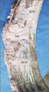
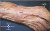
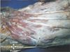
In 8 lower limbs (3 right, 5 left), this retinaculum was found distal to inferior extensor retinaculum extending across the width of the foot over the mid-foot region. Medially this retinaculum blended with plantar fascia covering the abductor hallucis muscle and laterally with the fascia over the base of 5th metatarsal (Fig. 1). Proximo-distally the retinaculum was about two fingers wide and covered the distal half of cuneiforms, cuboid and proximal thirds of metatarsals (i.e., over the tarsometatarsal joints). In two limbs (1 right, 1 left) the medial end of the retinaculum extended distally till the first metatarso-phalangeal joint (Fig. 2). The retinaculum had septations extending plantarwards to the metatarsals and the fascia over the dorsal interossei forming six osseofibrous compartments as shown in Fig. 3. The contents of the compartments (I to VI compartments) from medial to lateral side are shown in the Table 1. It was also noted that the septations as well as the retinaculum was thicker medially than laterally.
Partial retinaculum
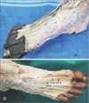
In 14 limbs (8 right, 6 left), the retinacular band did not extend completely across the width of the foot from its medial border to lateral border. It was limited to medial third of the foot (Fig. 4A, B). A single septum divided this band into two compartments corresponding to the first two compartments of the complete retinaculum and having similar contents. Attachments of partial retinacula are shown in Table 2. In one limb, an additional belly of extensor digitorum brevis (EDB) passed beneath the retinacular band along with extensor hallucis brevis (EHB), terminal branch of deep peroneal nerve (DPN), and the dorsalis pedis artery (DPA) (Fig. 5).
Discussion
In the present study, 22 specimens (44%) showed the presence of midfoot retinaculum, compared to 64.2% of 151 specimens by Kaneff and his colleagues (as quoted by Sarrafian) [4], which they called as retinaculum musculorum imum in man. Aktan Ikiz et al. [5] have also described a similar band crossing the DPN, DPA, and EHB in 61% of the 38 specimens. Lawrence and Botte [6] in their study of 17 specimens found a similar fibrous band although the prevalence was only 12%.
The presence of septations between the structures passing beneath the retinaculum has not been described in any study to the best of author's knowledge. Human foot is still undergoing evolutionary changes upon assumption of erect posture. The functional demands of bipedal gait with plantigrade foot are manifested in evolving musculature of human foot. Preventing bowstringing is imperative to ensure optimum action of long tendons on the distal joints of foot. This explains provision of additional retinaculum. The foot is more extensive in length medially. Provision of partial retinacula limited to medial aspect may be explained thus. Even the inferior extensor retinaculum is more expansive medially by splitting itself in a “y” like pattern. The retinaculum and septations probably enhance the stability of extensor tendons at the tarsometatarsal joints and prevent bow stringing.
The importance of knowing the presence of such a retinaculum lies in the fact that the retinaculum crosses DPA and the medial terminal branch of DPN and the EHB tendon, which are present within the compartment. This can give rise to entrapment neuropathy of the DPN if the nerve is stretched or compressed within it. This is more likely if an additional belly of EDB also passes through the second compartment as was observed in one limb in the present study.
Several studies have described anterior tarsal tunnel syndrome which is entrapment neuropathy of DPN beneath the inferior extensor retinaculum of the foot giving rise to either motor symptoms, sensory symptoms or both depending on the site of entrapment of the nerve [5678910].
Dellon [11] has described an area of “anatomic tightness” where EHB tendon crossed the DPN in the mid-foot region. In this area, the nerve is prone to entrapment in cases of injury in this area, high arched foot, swelling of the foot or a footwear with the band crossing this area [11]. Kanbe et al. [12] have also reported the entrapment neuropathy of DPN by the EHB. Presence of a retinacular band in this region, with septations, would thus predispose to entrapment of the nerve lying within and mimic symptoms of anterior tarsal tunnel syndrome.
To conclude, a partial or complete mid-foot retinaculum existed in the mid-foot region covering the tarsometatarsal joints in about half of study population. Functionally this retinaculum may prevent bowstringing of the extensor tendons, clinically it may predispose to entrapment of DPN mimicking anterior tarsal tunnel syndrome.
Limitations of the study is that sex of the specimen was not known. Hence, variations of retinaculum with respect to gender could not be assessed.
 XML Download