Journal List > Anat Cell Biol > v.50(2) > 1071890

Nayak, George, Mishra, Shetty, Sirasanagandla, and Padavinangadi: Surgical and radiological importance of a rare cysto-duodeno-colic peritoneal fold

Abstract

It is quite common to see abnormal peritoneal folds in the abdominal cavity. Some of them might compress or strangulate the viscera and others might determine the direction of the flow of peritoneal fluid, pus or blood. Many unusual clinically important peritoneal folds such as Ladd's band, cysto-gastro-colic fold, omento-cystic fold, and cysto-colic fold have been reported earlier. Knowledge of these folds is important for radiologists, gastroenterologists, and surgeons. We report an unusual cysto-duodeno-colic fold observed during our dissection classes. The fold was seen to compress the duodenum and colon. The fold extended from the descending part of the duodenum and the transverse colon to the gallbladder. It enclosed the entire gallbladder. A case similar to this has not been reported yet. It is important for the gastroenterologists and laparoscopic surgeons to be aware of this fold to avoid misdiagnosis and iatrogenic injuries.

Introduction

Peritoneum is the largest serous membrane in the body. It forms folds which suspend various viscera and support them. These folds are also the routes for the blood vessels to reach the viscera. Abnormal peritoneal folds are formed as a result of abnormal development and rotation of viscera or as a pathological process. Some of the reported abnormal peritoneal folds include Ladd's band [1], cysto-gastric fold [2], cysto-gastro-colic fold [3], omento-cystic fold [4], and cysto-colic fold [5]. We report a rare type of fold extending from descending part of duodenum and the right end of transverse colon to the Biologygallbladder, which can be called cysto-duodeno-colic fold.

Case Report

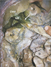
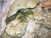
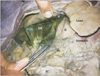
During dissection classes for first year medical students, we noticed an abnormal avascular peritoneal fold in a male cadaver aged approximately 60 years. The fold extended from the descending part of the duodenum and right end of the transverse colon to the gallbladder (Figs. 1, 2, 3). It enclosed the entire gallbladder and appeared as if it was pulling the gallbladder in downward direction. The tension of the fold had possibly compressed the descending part of the duodenum and the right end of the transverse colon. This was evident as the first part of duodenum and the right colic flexure were dilated. The transverse colon at the area of attachment of the fold was narrowed.

Discussion

Abnormal peritoneal folds are observed frequently during surgery and cadaveric dissections. Most of the abnormal folds are asymptomatic and go unnoticed throughout life. Some of them might be involved in harbouring disease processes or serve as channels for the spread of malignancies. Another problem associated with such abnormal peritoneal folds is compression and obstruction of the hollow viscera. Low et al. [6] have reported the radiographic observation of a fold compressing the duodenum. Compression of proximal jejunum by a congenital band has been reported by Liu et al. [7]. Reports are also available on the peritoneal folds connecting the gallbladder to greater omentum, lesser omentum and right kidney [48]. When the peritoneal folds enclosing the gallbladder are very long, they might result in a condition known as “Floating gallbladder” and result in torsion or volvulus formation [910].
In the current case, the peritoneal fold extended from descending part of duodenum and right end of transverse colon to the gallbladder. Its compressive/traction effect on the gut was evident in the form of dilation of the first part of duodenum and the right colic flexure. The fold had also caused a stricture on the wall of the transverse colon. This fold of peritoneum might cause adverse effects in the normal functioning of the gallbladder and the gut. It might mislead the gastroenterologist and radiologist in their diagnosis. To a surgeon, it might cause confusions during laparoscopic cholecystectomy. Hence, a prior knowledge of possibility of existence of a cysto-duodeno-colic fold is of tremendous importance to the surgeons, gastroenterologists and radiologists.

Figures and Tables

Fig. 1

Dissection of the upper abdomen showing the cysto-duodeno-colic fold. AC, ascending colon; CDCF, cysto-duodeno-colic fold; D, first part of the duodenum (note the dilation); GB, gallbladder enclosed in the fold; RCF, right colic flexure (note the dilation); SI, small intestine; TC, transverse colon.

acb-50-159-g001
Fig. 2

Closer view of the cysto-duodeno-colic fold. C, compressed (narrow) part of the transverse colon; CDCF, cysto-duodeno-colic fold; D, first part of the duodenum (note the dilation); GB, gallbladder enclosed in the fold; RCF, right colic flexure (note the dilation); SI, small intestine; TC, transverse colon.

acb-50-159-g002
Fig. 3

Side view of the cysto-duodeno-colic fold. CDCF, cysto-duodeno-colic fold; D, first part of the duodenum (note the dilation); GB, gallbladder enclosed in the fold; TC, transverse colon.

acb-50-159-g003

References

1. Mongardini M, Moschella CM, Schillaci F, Cola A, Fanello G, Benedetti F, Maturo A, Pappalardo G. Strangulated abdominal herniation by Ladd's band. G Chir. 2005; 26:207–209.
2. Nayak SB, Shetty SD, Surendran S, Jetti R, Kumar N, Sirasanagandla SR. Double gallbladder completely enclosed in a cystogastric fold of peritoneum. Anat Cell Biol. 2014; 47:132–134.
3. Pamidi N, Nayak S, Vollala VR. Cystogastrocolic fold and associated atrophy of the gallbladder. Singapore Med J. 2008; 49:e250–e251.
4. Nayak SB, George BM, Mishra S, Ashwini LS, Marpalli S. Omento-cystic peritoneal fold and rudimentary quadrate lobe: a case report. OA Case Rep. 2013; 2:46.
5. Mehta V, Arora J, Suri RK, Kumar H, Gupta V, Rath G, Das S. Clinical relevance of a cysto-colic peritoneal band found incidentally during cadaveric dissection. Clin Ter. 2010; 161:153–154.
6. Low VH, Davis SJ, Yoong MF. Anomalous peritoneal folds of the duodenum: a normal variant simulating disease. Australas Radiol. 1992; 36:135–136.
7. Liu C, Wu TC, Tsai HL, Chin T, Wei C. Obstruction of the proximal jejunum by an anomalous congenital band: a case report. J Pediatr Surg. 2005; 40:E27–E29.
8. Nayak SB. Abnormal peritoneal fold connecting the greater omentum with the liver, gallbladder, right kidney and lesser omentum. Bratisl Lek Listy. 2009; 110:736–737.
9. Tzardinoglou E, Prousalidis J, Apostolidis S, Katsohis C, Aletras H. Hanging noncalculous gallbladder. HPB Surg. 1996; 9:137–139.
10. Shaikh AA, Charles A, Domingo S, Schaub G. Gallbladder volvulus: report of two original cases and review of the literature. Am Surg. 2005; 71:87–89.
TOOLS
Similar articles