

 PDF
PDF ePub
ePub Citation
 Print
Print


Introduction
The conventional concept of angiogenesis stated that endothelial cells initiate a process to uncover the basement membrane which later is digested by enzymes to leave some gaps. Through these gaps endothelial cells migrate and divide to form loops and tubes. A new basement membrane is then secreted and the open lumen of the new vessel communicates with that of the mother vessel [1]. Whether the same mechanism operates in human lymphatic development remains to be elucidated.
The choice of a suitable material for the study of human lymphatic development is difficult. In this study, surgically excised human mature cystic ovarian teratomas were chosen. Patients' consent and institutional review board permission were obtained.
Materials and Methods
Tissue blocks from solid areas of five specimens were excised and embedded in paraffin, 5 µm sections were mounted and stained with hematoxylin and eosin.
For electron microscopy, the tissues were fixed in 3% phosphate buffered glutaraldehyde and 1% osmium tetroxide, dehydrated in ascending ethanol, passed through propylene oxide, embedded in Spurr's resin and cut with an ultramicrotome. Lymphatics were identified in semi-thin sections and trimmed for ultra-thin sectioning, then stained with uranyl acetate and lead citrate and examined using a Jeol electron microscope (Tokyo, Japan), magnification ×100,000.
Results
Light microscopy
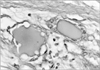
Areas of eosinophilic amorphous extracellular substance surrounded by several cells were observed. The nuclei had dispersed chromatin and did not appear to be at any stage of mitosis. The cytoplasm was scarce and extended in the form of cellular processes. Some processes were in conjunction with the cells lining the extracellular substance, while others projected away and formed a meshwork containing a less dense matrix. These findings were interpreted as a starting point of developing lymphatic capillaries lined by mesenchymal cells (Fig. 1).
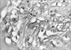
In one section, the scalloped eosinophilic substance was surrounded by several cells similar to those shown in Fig. 1, but with thinner nuclei and less numerous cytoplasmic processes, especially those forming a meshwork nearby (Fig. 2). This vessel has been interpreted as a more developed lymphatic because of its continuous lining of mesenchymal cells.
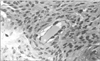
In a longitudinal section of a similar vessel, zones lined by slender cytoplasmic processes were intermingled with zones lined by a thicker cytoplasm harboring the cell nuclei. The outgoing cytoplasmic processes were fewer and shorter. The general appearance of a well formed lymphatic was observed in Fig. 3.
Electron microscopy
In the early stages of lymphatic vessel formation, the lining mesenchymal cells had large nuclei, scarce perinuclear cytoplasm and multiple thin cytoplasm processes and few organelles, as usual for undifferentiated cells.
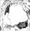
In the more advanced stages of vessel formation, the lining cells showed microvesicles, in the form of caveolae or pinocytotic vesicles, overlapping intercellular junctions as well as tight junctions. The cytoplasm had more developed ribosomes and mitochondria. The nuclei were flatter and had a chromatin pattern similar to that of endothelial cells. Interdigitating junctions were only seen at a very late stage and only in fully developed lymphatics, which were scarce in this type of specimen (Fig. 4).
Discussion
Light and electron microscopy describe “endothelial-like” structures in lymphatics [2]. This resemblance may mean that these cells look like endothelium but do belong to some other category of cells. This study suggests that this resemblance is simply because the cells are in a developmental stage, before differentiating into endothelial cells.
There is a consensus that lymphatics should be lined by endothelium. The present study investigated developing lymphatics, where typical endothelial lining was not always observed. Instead, the amorphous eosinophilic content was considered diagnostic.
Immunocytochemistry is widely used to detect specific surface antigens of endothelial cells. Utilizing this method, Conzonieri et al. [3] have shown focal expression of factor IX related antigen by putative endothelial cells in some cysts.
Klika et al. [4] found that mesenchymal cells form part of the wall of the primitive lumphatics of the epicardium of check embryo. Al-Jomard and Scothorne [5] demonstrated that lymphatics in rat liver develop from in-situ differentiation of mesenchymal cells into endothelial cells. These findings are further evidence to the conclusion of this study that the lymphatic endothelium in human mature cystic ovarian teratoma is of mesenchymal origin.
 XML Download