

 PDF
PDF ePub
ePub Citation
 Print
Print


Introduction
We recently describe the morphology of the synovial tissues in the cricothyroid (CT) joint in Japanese elderly [1]. We find that the CT joint is characterized by drastic degeneration of the synovial tissues, including exposure of the external ligament fibers to the joint cavity due to destruction of the capsule. However, despite this severe degeneration, synovial macrophages are usually few in number. In contrast to the CT joint (which is almost flat), the cricoarytenoid (CA) joint is similar to a saddle joint, such as the finger joints. The CA joint has been one of the major focuses of anatomical studies of phonation, perhaps to a greater degree that the CT joint, and for elderly individuals a lot of detailed information exists regarding loss of elastic cartilage and the subsequent change to bone tissue in the arytenoid [23456]. Likewise, roughness and fibrillation of the surface articular cartilages have been reported, especially in marginal areas [7]. In contrast, it has been considered that synovial tissue, including the capsule, remains stable with increased age [5]. However, to our knowledge, no previous study has identified synovial macrophages lining the CA joint cavity, even though these cells are one of the usual components of synovial tissues in the musculoskeletal system [89]. Therefore, we conducted the present study to compare the morphology of the synovial tissue between the CA and CT joints.
Materials and Methods
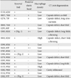
The study was performed in accordance with the provisions of the Declaration of Helsinki 1995 (as revised in Edinburgh 2000). We examined 18 donated cadavers (15 men and 3 women) ranging in age from 62 to 97 years, with a mean age of 85 years. The cause of death had been ischemic heart failure or intracranial bleeding. These cadavers had been donated to Tokyo Dental College for research and education on human anatomy, and their use for research did not require approval by the university ethics committee. The donated cadavers had been fixed by arterial perfusion with 10% v/v formalin solution and stored in 50% v/v ethanol solution for more than 3 months. From each cadaver, we prepared one tissue block that included the CA joint and the other small structures around the joint. Thus any left/right difference was not examined. The sectional plane was sagittal or nearly sagittal (tilted sagittal). The specimens were decalcified by incubating them at 4℃ in 0.5 mol/l EDTA solution (pH 7.5, decalcifying solution B, Wako, Tokyo, Japan) for 7–14 days. Half of the 18 cadavers (Table 1) overlapped with our recent study of the CT joint [1].
After performing routine procedures for paraffin-embedded histology, semiserial sections, i.e., adjacent or near 9–12 sections, were prepared at almost 0.5-mm intervals. Two of the sections were stained with hematoxylin and eosin and elastica-Masson (a variation of Masson-Goldner staining [1011]), respectively. The other sections were used for immunohistochemistry. The primary antibodies used were (1) mouse monoclonal anti-bovine alphaelastin (1:20, ab9519, Abcam, Cambridge, UK), (2) rabbit polyclonal anti-human factor VIII-related antigen (von Willebrand factor) (1:100, Dako IR527, Dako, Glostrup, Denmark),(3) mouse monoclonal anti-human CD68 KP1 (1:100, M0814, Dako), (4) rabbit polyclonal anti-human IgM (1:100, Dako N1509, Dako), (5) mouse monoclonal antihuman CD79a (1:40, Dako M7050), (6) mouse monoclonal anti-human CD3 (1:100, Nichirei 413591, Tokyo, Japan), and (7) mouse monoclonal anti-human CD8 (1:100, Dako N1592, Dako). Antigen retrieval with microwave treatment (500 W, 15 minutes, pH 6) was performed for antibodies Nos. 2–7, while trypsin treatment was used for for antibody No. 1. The secondary antibody (incubation for 30 minutes, 1:1,000, Histofine Simple Stain Max-PO, Nichirei) was labeled with horseradish peroxidase (HRP), and antigen-antibody reactions were detected by the HRP-catalyzed reaction with diaminobenzidine (incubation for 3–5 minutes, Histofine Simple Stain DAB, Nichirei). Counterstaining with hematoxylin was performed on the same samples. A negative control without the first antibody was set up for each of the specimens.
Although elastica Masson staining is not widely used, it colors elastic fibers clear black in contrast to a bright green color for collagen fibers. Among the above antibodies, that against CD68 was used for identification of macrophages, those against CD3 and CD8 for T lymphocytes, those against IgM and CD79a for B lymphocytes, and that against factor VIII for blood capillaries. There are many antibodies for detection of lymphocytes, such as anti-CD4 antibody for T lymphocytes, but most of them cannot be applied to long-preserved specimens from donated cadavers [12]. Observations and photography were usually performed with a Nikon Eclipse 80 (Nikon, Tokyo, Japan).
Results
Joint cartilage
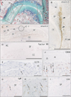
Both the arytenoid and cricoid cartilages were ossified in all specimens. The CA joint surface of the arytenoid cartilage was consistently concave, in contrast to the convex shape of the cricoid joint surface. The joint cartilage (hyaline cartilage) was usually larger in the arytenoid or upper side than in the cricoid or lower side in the present sagittal or tilted sagittal sections (Figs. 1,2,3). The upper surface of the CA joint tended to extend medially over the medial margin of the cricoid cartilage. Cartilage thinning was seen in three of the 16 specimens and roughness was evident in another three specimens (Fig. 1A). In this series we did not find any cartilage defect that resulted in exposure of bone tissue to the joint cavity.
Capsule and other synovial tissues
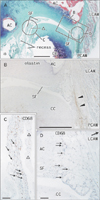
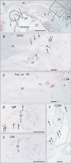
The CA joint capsule was thin and contained few elastic fibers (Figs. 1C, 2B). The lateral and posterior aspects of the CA joint were covered by the lateral and posterior CA muscles, respectively (Figs. 1A, 2A, 3A). However, in association with increased fatty tissues in and around the muscle, thin loose tissue was usually (15/18) interposed between the muscle and the capsule (Figs. 2A, B, 3A, C). This loose tissue contained a few nerves and vessels. The covering fascia of the posterior CA muscle was often (10/18 specimens) thick and the maximum thickness reached 0.1 mm. We referred to this as the "posterior band" (Table 1), and it was found to contain abundant elastic fibers (Figs. 1B, 2B). When the elastic fiber-rich fascia was attached to the posterolateral aspect of the joint capsule (Fig. 1B), the capsule itself was difficult to discriminate from the fascia. In contrast, the medial and anterior aspects of the CA joint faced a large area of loose tissue that was continuous with the laryngeal submucosal tissue. Therefore, there was never any definite supporting structure in the anterior and medial aspects of the CA joint. The marginal parts of the CA joint cavity were usually enlarged to provide recesses (Figs. 2A, 3A).
Synovial folds were consistently present in the CA joint: the short posterior fold was a triangular mass, while the long anterior fold was composed of multiple belt- or tongue-like thin folds (Figs. 1A, 2A, 3A). The synovial fold tended to cover an area of the joint surface showing cartilage roughness (Fig. 1A). The capsule and synovial folds usually contained multiple capillaries (Figs. 2C, 3C). CD68-positive macrophages as well as lymphocytes were usually sparse or absent along the joint cavity: 0–10 cells per 100 µm2 in section (Figs. 1E–I, 2C, D, 3B, D–F). However, in two specimens (from males aged 82 and 93 years), the synovial macrophage density was relatively high (10–20 cells per 100 µm2): one of these 2 specimens carried well developed folds, while another did not (Table 1). We sometimes observed round or oval large cells that were positive for CD3 (Fig. 1H), but these did not appear to be usual T lymphocytes on the basis of morphology. Overall, in most specimens, we did not find any evidence of synovitis despite the fact that joint cartilage degeneration was sometimes present.
Comparison with the CT joint
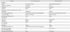
Nine of 30 cadavers from which specimens of the CT joint had been obtained for our recent study [1] were used for the present comparative study of the CA joint (Table 1). One of these specimens (from an 82-year old man) showing a high density of macrophages dislayed multiple signs of degeneration in the CT joint. However, another specimen (from a 94-year-old man) in which the CT joint had been obliterated by elastic fiber-rich tissues (i.e., showing the most severe degeneration) did not exhibit specific or severe degeneration in the CA joint. Overall, we did not find any clear correlation between CT joint degeneration and CA joint degeneration. Observations of the CA joint are summarized in Table 2 for comparison with the CT joint. In short, the CA joint did not show drastic degenerative changes such as obliteration of the joint cavity or exposure of the ligament component fibers to the joint cavity.
Discussion
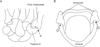
Although a site-dependent difference in supportive structures was commonly evident in the CT joint, the CA joint was characterized by weakness or even absence of such structures. Lacking any ligament, the anterior and medial aspects faced loose tissue that was continuous with the laryngeal submucosal tissue. A photo presented by Casiano et al. [5] suggested that this medial weakness is likely to be present in young individuals. Indeed, the lateral and posterior aspects were covered by the lateral and posterior CA muscles. However, loose tissue was often interposed between the thin capsule and muscle, possibly due to age-related degeneration. In contrast to the definite capsular ligaments of the CT joint, i.e., the anterior band and posterior mesh of elastic fibers [1], the so-called CA ligaments might be restricted posteriorly and correspond to the posterior band of elastic fibers. The CA joint is similar to a saddle joint with high congruity (Fig. 4B). Moreover, in all of the specimens examined, synovial folds seemed to increase and maintain this high congruity. Using Indian ink pin-prick assessment, Kahn and Kahane [7] reported marginal roughness and fibrillation of the CA joint surface: this marginal lesion may correspond to sites covered by the synovial folds. Notably, a typical saddle joint, i.e., the carpometacarpal joint of the thumb (Fig. 4A), is characterized by looseness of the ligaments and capsule to allow a wide range of movement including slight rotation at the neutral position [1314]. Therefore, the weak ligament of the CA joint also appears to allow rotation and sliding depending on the actions of the laryngeal muscle.
Although perhaps an oversimplification, vibration of the vocal fold is likely to be conducted to the arytenoid, and via the cricoid to the thyroid cartilage. This hypothetical conduction route is interposed by the CT and CA joints. Does age-related degeneration correlate between the two laryngeal joints? In saddle-like finger joints, Nakamura et al. [15] reported that cartilage degeneration shows radialside dominance. In the ankle of elderly individuals, Hirose et al. [16] described that the sites of cartilage degeneration were correlated between the talocrural and subtalar joints. Progressive degeneration in these two joints would likely be correlated because, during gait, a limitation of motion range in one joint can be compensated, to some extent, by the other. However, in the CT and CA joints, there seemed to be a critical difference in the mechanism to minimize vibration: abundant elastic fibers in the thick ligaments would likely avoid injury due to vibration in the CT joint [1], while in the CA joint, joint congruity would seem to be more important for function than a stabilizing effect of ligaments. The strong laryngeal muscles inserting to the arytenoid may absorb part of this vibration. Vibration may simply be conducted along a straight line from the upper arytenoid to the lower cricoid, in contrast to a change in the vector between the cricoid and the inferior cornu of the thyroid cartilage. When considered together with the well developed synovial folds containing few elastic fibers, any influence of vibration in accelerating degeneration appears to be much less pronounced in the CA joint than the CT joint.
 XML Download