

 PDF
PDF ePub
ePub Citation
 Print
Print


Introduction
The supratrochlear foramen (STF) is an important and relatively common anatomic variation in the lower end of the humerus in humans. A thin, transparent plate of bone known as supratrochlear septum varying from 0.5 mm to 1 cm in thickness, which is lined in fresh state by the synovial membrane of elbow joint separates the olecranon and coronoid fossae. This septum may contain several perforations and in some cases may become perforated to form an aperture known as ‘supratrochlear aperture’ or ‘supratrochlear foramen’. It was first described by Mekel in 1825 [1]. STF has been designated by a variety of names such as intercondylar foramen, olecranon foramen and epitrochlear foramen [23]. According to Hirsh (1927), the thin plate of bone between the olecranon and coronoid fossa is always present until the age of seven years, after which the bony septum occasionally becomes absorbed to form the STF [4]. Individuals with this variation may be able to overextend the elbow joint [5]. It has been described in hyenas, dogs, and other primates [6]. The septal apertures have not been observed only in humans but in most mammalian species, with a slightly greater prevalence amongst the old and new world apes [7]. Darwin considered this foramen in humans, as one of the characteristics to show the evolutionary aspect of humans from primates [8].
T-Box (TBX) genes may play a role in STF formation. Genes of the TBX family control the synthesis of proteins known as TBX proteins that are crucial for the development of the limbs and the heart in utero [9]. This concept was furthered by Govoni's work which postulated that TBX genes effect postnatal development of the limbs [10]. The STF was a hitherto unimportant descriptive feature of the humerus. Based on orthopaedic surgical experience, it has been noted that STF is associated with a narrow intramedullary cavity [11]. In the paediatric age group the supracondylar fracture is of common occurrence [12]. This is treated by intramedullary nailing which may be compromised by the presence of this aperture. The retrograde nailing is done through the medial and lateral epicondyles or only through the lateral [13]. Nerve injuries become pertinent. Therefore its presence is of utmost importance in planning the nailing of fractures of the lower end of the humerus [1112]. During radiological evaluations of the humerus, presence of STF may result in erroneous interpretation as pathological lesions or cysts [13]. The present study on south Indian population is an attempt to highlight the incidence, morphological features, and clinical importance of STF which may be beneficial to orthopedic surgeons and radiologists.
Materials and Methods
The present study was conducted on dried adult human humeri of unknown sex and free of pathological changes, of south Indian origin, in the Department of Anatomy, Sri Siddhartha Medical College, Tumkur, Karnataka, India. The presence of a STF was noted; its shape was observed and divided into three types (oval, round, and irregular). The transverse and vertical diameters of the STF were measured using a vernier caliper. In bones where the foramen was absent the translucency of the septum between the coronoid and the radial fossa was noted by placing the lower end of the humerus against the X-ray view box and, to confirm that, the X-ray of the lower end of the humerus (postero-anterior view) was taken. All observations were recorded in a tabular form.
Results
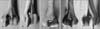
Out of the 142 humeri studied, 72 humeri (50.7%) were right sided and 70 humeri (49.2%) were left sided. In these 142 humeri, 38 humeri (26.7%) showed the presence of STF. In these 38 humeri with STF, 16 (22.22%) were right sided and 22 (31.43%) were left sided. The incidence of STF was more on the left side than the right side (Table 1). The majority of STF were round shaped in 47.37% (Fig. 1), followed by oval shaped in 42.11% and 10.53% were irregular in shape (Table 2). The STF was absent in 36 humeri (25.35%) and 68 humeri (47.89%) showed the translucency of septum (Table 3). The mean transverse diameter on right side was 4.50±3.183 mm and 3.32±3.222 mm on left side. The mean vertical diameter was 3.88±2.391 on right side and 3.6±3.532 mm on left side (Table 4). The side differences of the diameters were not statistically significant.
Discussion
Foramina are defined as conduits for vessels and nerves, while apertures are merely openings in bones [14]. No anatomical structure is known to pass through the STF and hence does not qualify to be one by definition. Its aetiology and function are largely obscure. Two theories for the formation of this foramen have been proposed. A more solid hypothesis suggests that the septal apertures are purely mechanically induced (overgrown olecranon or coronoid process, a wider range of flexion and extension at the elbow joint) and it may be worth viewing it as part of the anatomical and mechanical relationship between humerus and ulna during normal flexion and extension of the elbow [14]. This theory is also supported by the views of some researchers that statistical data that humeral septal apertures were commonly presented in the Neolithic and prehistoric types but are relatively rare in late Europeans and Hirsh predicted: if it can be proven that there is any relationship between the presence of the foramen and an increase in the flexion and extension amplitude at the elbow joint, its significance would be apparent. It would then mean that there has been an adaptation to functional needs and that, on the whole modern man has lost to a certain degree the range of these particular motions possessed by his prehistoric ancestors and animals in whom this foramen is more or less constant finding and that this character is in recession and will eventually disappear. Studies have shown that the humerus is not perforated in the embryonic stage. The foramen usually develops during adolescence, or more frequently adulthood due to incomplete ossification, due to intralamellar space enlargement and gradual septum absorption. Overall, studies have shown that septal apertures were more common among ancient human populations, specially in late Europeans. The frequency of humeral septal apertures appears to have decreased considerably since the Paleolithic and even the Neolithic times. Since pressure from the olecranon should act as strengthening factor, it has been suggested that STF may be caused by insufficient blood supply [15]. Therefore its presence can be attributed to the functional development of the elbow joint and its variable shapes to the morphometry of the articulating surfaces. Its left side frequency can be explained on the basis of handedness. It is more common on the non-dominant left hand as the dominant right limb has more robust bones [1516]. The prevalence of STF in the present study is 26.7%. The prevalence of the STF in the human population varies from 0.3% to almost 60% worldwide (Table 5). In a study conducted on Libyas by Macalister, the prevalence was 57% [17]. With no other obvious cause, the observed communication between the olecranon fossa and coronoid fossa seems to be a rather unusual congenital irregularity. Congenital deformities of bilateral occurrence might reflect the presence of a particular, though very rare genetic defect, since an Online Mendelian Inhritance in Man (OMIM) database searchdid not reveal any disorder with a clinical evaluation corresponding to distal upper limb communication. Such congenital deformity might be due to an overgrown olecranon or coronoid process, or it could be related to collagen fibre elasticity causing loose joints. Loose joints commonly overextend and this could cause an erosion of the thin bone septum between the fossae. In this way laxity, which is more often associated with women, may justify this kind of bilateral trauma as well as its increased occurrence in females. The prevalence was 0.3% in a study on Greeks by Papaloucas et al. [18]. In the present study, the prevalence of the STF was higher on the left side. This result was consistent with earlier studies on Koreans, Egyptians, and Eastern Indians [21920]. The prevalence of STF on various populations by different authors is shown in Table 5 [21222324]. Hrdlicka reported that it was more common in the Australians, the yellow-browns in general (exclusive of the Lapps), and in the Negro, than it is in the European whites [25]. A study conducted by Benfer and McKern [26] showed that the prevelance of STF in Americans was 6.9%. In the present study, majority of STF (47.37%) were round. Next to round, oval shape being common was in 42.11% and irregular in 10.53%. Veerappan et al. [27] observed oval shape in 42.85%, round shape in 37.71%, triangular shape in 14.28% and sieve like in 7.14% in their study. Translucency of the septum was observed in 47.89% of specimens and the STF was opaque in 25.35% in present study. Nayak et al. [12] found the translucency in 56.7%, Veerappan et al. [27] observed such an incidence in 50%. In the present study, the mean transverse diameter of the STF was 3.32 mm and 4.50 mm, on left and right sides respectively while the vertical diameter was 3.86 mm and 3.88 mm on left and right sides respectively. This result was consistent with a study by Mathew et al. [28]. The side differences of the diameters were not statistically significant in the present study. The results obtained in the present study were compared with that of other authors like Nayak et al. [12], Krishnamurthy et al. [24], and Veerappan et al. [27] (Table 6).
Supracondylar fractures account for approximately 17% of pediatric trauma. Retrograde intramedullary nailing forms the mainstay of treatment [29]. As it is said that STF is associated with a narrow medullary cavity antegrade route may be preferred. So, knowledge of its presence helps decide the surgical course of action. STF appears relatively radiolucent and may commonly be confused with osteolytic or cystic lesion. STF shows wide racial differences and hence can be used as a parameter in racial identification. Its link as an evolutionary trait can be used for dating specimens by the anthropologist [30].
In conclusion, the STF is an entity familiar to the anthropologist and the anatomist but has not received much notice from the clinician, evidenced by it cursory mention or not at all in most textbooks of clinical practice. As it is not a passage for any neurovascular structure, we propose that the term supracondylar aperture would be more apt terminology. Its existence is important to the orthopaedician in the preoperative planning of nailing fractures of the distal humerus and to the radiologist for differentiating it from an osteolytic or cystic lesion. The anatomical knowledge of STF is beneficial for anthropologists, orthopaedic surgeons and radiologists in day to day clinical practice.


 XML Download