

 PDF
PDF ePub
ePub Citation
 Print
Print


Introduction
Cephalic vein is one of the superficial veins of the upper limb. It runs along the lateral border of the forearm and the arm, passes through the delto-pectoral groove, pierces the clavipectoral fascia at the delto-pectoral triangle and opens into the axillary vein. Variations of cephalic vein are rare. Its variations include its total absence, termination into subclavian vein, and abnormal communication with the external jugular vein [12345]. In very rare cases, it crosses superficial to the clavicle and terminates into external jugular vein. This type of termination is called persistent jugulocephalic vein because it is derived from an embryonic channel that connects the cephalic vein with the external jugular vein [6]. We found one such persistent jugulocephalic vein during our dissection classes and the aim of this case report is to discuss the clinical implications of this rare variation. The knowledge of this variation may be very useful to the radiologists, orthopedic surgeons, and plastic surgeons.
Case Report
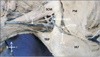
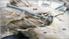
During routine dissection classes for medical undergraduates, we observed a rare termination of cephalic vein of the right side in an adult male cadaver aged approximately 70 years. The variation was unilateral. The cephalic vein had a normal origin and course till it reached the delto-pectoral triangle. Upon reaching the delto-pectoral triangle, instead of piercing the clavipectoral fascia and terminating into the axillary vein, it ascended in front of the lateral part of the clavicle and terminated by opening into the external jugular vein. It received a large muscular vein from sternocleidomastoid muscle just before its termination. It also gave a communicating vein at the delto-pectoral triangle, which pierced the clavipectoral fascia and opened into the axillary vein. External jugular vein received the posterior external jugular vein and terminated into the subclavian vein. These variations are shown in Figs. 1 and 2. The diameters of the veins observed were as follows: jugulocephalic vein, 0.3 cm; external jugular vein, 0.6 cm; axillary vein, 1 cm; cephalic vein, 0.5 cm; basilic vein, 0.4 cm; and communicating vein, 0.3 cm. The other veins in this case did not have any variations.
Discussion
Cephalic and external jugular veins are the two superficial veins which are used in many clinical and surgical procedures. A thorough knowledge of their variations can contribute greatly to the success of any radiological or surgical procedures. During embryonic development, the preaxial vein of the upper limb develops into cephalic vein and the postaxial vein develops into basilic vein. During early stages of development, the cephalic vein drains into a venous plexus of neck. The external jugular vein develops from this plexus. Thus, in the early fetal life, cephalic vein is a tributary of external jugular vein. In a later stage of development, the cephalic vein establishes a connection with the axillary vein, after which it loses its connection with the external jugular vein. If it fails to lose connection with the external jugular vein, then it is called persistent jugulocephalic vein [36789]. Cephalic and external jugular veins are generally used for central venous catheterizations. Many device implanters prefer cephalic vein cut down for the placement of defibrillator leads instead of directly puncturing the axillary or subclavian veins to avoid the risk of pneumothorax, subclavian crush, and other complications. However, when there is a persistence of jugulocephalic vein, the catheterization may not be successful [10]. In a study conducted by Loukas et al. [1], the cephalic vein terminated into subclavian vein after crossing anterior and superior to the clavicle in 0.2% of cases. In these cases, it gave a communicating branch to the external jugular vein. Nayak and Soumya [11], have also reported a communication between cephalic and external jugular veins superficial to the clavicle. But in that case, the cephalic vein terminated into the axillary vein by passing below the clavicle. In the current case, the cephalic vein terminated into external jugular vein by passing superficial to the clavicle and gave a communicating branch to the axillary vein. Supraclavicular course of cephalic vein is very rare and available reports indicate that it occurs only in 0.2% cases [12]. Cephalic vein with supraclavicular course must be avoided during pacemaker implantation due to the risk of skin erosion or lead fracture. Świętoń et al. [13] suggest that supraclavicular course of cephalic vein might significantly affect the first time or repeated placement of cardiac implantable electronic device. According to Wysiadecki et al. [6] persistence of jugulocephalic vein increases the risk of complications during the clavicular fractures, cephalic vein catheterization, or head and neck surgery. Lin et al. [14] recommend external jugular vein cut-down in cases of failure of cephalic vein cut-down procedures. Apart from the above said procedures, the persistence of jugulocephalic vein is of cosmetic concern also, and can be safely excised for the same reason [15].
Variations of cephalic vein are rare. Among its known variations, persistence of jugulocephalic vein is extremely rare. Knowledge of the same is quite useful to orthopedic surgeons, plastic surgeons, head and neck surgeons, cardiovascular surgeons and more importantly, to the radiologists. Possibility of persistence of jugulocephalic vein has to be kept in mind during catheterizations, placement of cardiac devices and while treating the fractures of clavicle.
 XML Download