

 PDF
PDF ePub
ePub Citation
 Print
Print


Introduction
The estimation of stature from unknown human skeletal remains is one of the most significant stages of establishing personal identity in forensic investigations. As such, several researchers have investigated the possibility of estimating stature from different bones of the human skeleton [123456789101112]. Normally, the research is designed to acquire specific measurements from the skeletal remains and the data is analyzed using linear regression, from which stature-estimating formulae are derived. The regression formulae based on the length of the extremity long bones, particularly the femur, are considered the best estimators of stature [8]. However, such regression formulae cannot be readily applied since postmortem femora obtained in forensic investigations are usually incomplete (fragmented) as a result of decomposition and decay. Thus, it has become necessary to estimate stature from fragmentary femora.
The estimation of stature from fragmentary femora can be carried out by using direct and indirect methods [13]. The latter method involves initial estimation of the length of the femur from measurements of fragments of the femur followed, by the estimation of stature from the estimated femoral length (FL) using regression analysis [13]. Several regression formulae have been suggested for estimating FL from measurements of different anatomical portions of the femur [8141516].
Though useful, these regression formulae vary in different populations as a result of genetic and geographical factors. Thus, a single regression formula cannot be used worldwide. To date, no study has established such regression formulae using Ghanaian skeletal remains. The current study, therefore, seeks to derive preliminary regression formulae for the reconstruction of FL from fragmentary femora of Ghanaians.
Materials and Methods
The skeletal materials used in the current study comprised 50 adult dry femora obtained from the teaching skeletal collection at the Department of Anatomy, Kwame Nkrumah University of Science and Technology, Ghana. Demographic characteristics such as age, ethnicity and occupation of the individual bones were unknown. All the femora were devoid of any gross abnormalities that could preclude accurate measurement. Six measurements were acquired from different portions of the femur using a sliding caliper (Shanghai Medfriend Co. Ltd., Shanghai, China). The measurements were as follows:
(1) Vertical head diameter (VHD): maximum vertical diameter of head between the highest and the lowest point of the head.
(2) Transverse diameter of head (TDH): the maximum diameter from medial to lateral perpendicular to VHD.
(3) Bicondylar breadth (BB): distance between the two most laterally projecting points on the condyles.
(4) Epicondylar breadth (EB): distance between the two most laterally projecting points on the epicondyles.
(5) Sub-trochanteric transverse diameter (STD): distance between medial and lateral surfaces at the proximal end of the diaphysis just below the lesser trochanter.
(6) Sub-trochanteric anterior-posterior diameter (SAPD): distance between anterior and posterior surfaces at the proximal end of the diaphysis, measured perpendicular to the STD.
In addition, FL was measured as the distance from the most superior point on the head of the femur to the most inferior point on the distal condyles using a locally designed osteometric board, with attached tape measure (The Perfect Measuring Tape Company, Toledo, OH, USA).
All measurements were performed by one observer and recorded to the nearest 0.1 mm. Before the actual measurements, all measurements were taken twice on 20 femora at different time periods. Intra observer differences between these two measurements were compared using a paired t test. A non-significant result (P>0.05) indicated that the two measurements were not very different and were reproducible without significant intra observer errors. The relationship between FL and other femoral measurements was assessed using Pearson correlation analysis. In addition, linear regression formulae were derived (from all the significant correlations) for prediction of FL. The estimated FL was compared with the measured FL by calculating the mean discrepancies between the measured and predicted data (i.e., standard error of estimate, SEE). All statistical analyses were performed using SPSS version 20 (IBM Corp., Armonk, NY, USA). A value of P<0.05 was considered statistically significant.
Results
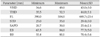
Table 1 shows statistical descriptive summaries of all the measurements taken from the femur. Table 2 shows the linear regression analysis of the relationship between FL and other measurements (fragments) of the femur. The table also shows the correlation coefficients (R), coefficients of correlation (R2), regression formulae for estimating FL from different femoral measurements, mean difference between the estimated FL and measured FL (i.e., SEE), and P-values. All the six parameters showed significant positive correlations with FL (P<0.01). The correlation coefficients ranged from moderate to high (0.580–0.819) with STD and BB producing the highest and least values, respectively. The mean discrepancies between the estimated and measured FL ranged from 13.66 mm and 19.28 mm with STD and BB producing the least and largest discrepancies, respectively.
Discussion
In this study, six measurements were acquired from different anatomical portions of the femur, and assessed for prediction of FL using linear regression analysis. The measurements include VHD, TDH, BB, EB, SAPD, and STD. The results indicated significantly moderate-to-high correlations (r=0.580–0.819) between each of these measurements and the FL. The error estimates of the regression formulae were relatively low (i.e., SEE, 13.66–19.28 mm), suggesting that the discrepancies between actual and estimated stature were relatively low. STD emerged as the best estimator of FL when compared with the other assessed measurements of the femur.
These results are consistent with other studies, which have shown that portions (fragments) of the femur can be used to estimate the length of the femur [81415]. The correlation coefficients and SEE of comparative measurements in the current study are quite similar to those reported by Chandran and Kumar (i.e., R=0.618–0.811, SEE, 8.88–11.95 mm) [14].
Most studies conducted on the reconstruction of FL from femoral fragments have assessed fragments of the epiphyses (ends of the femur) [814]. The most commonly assessed parameters include VHD, vertical neck diameter, upper breadth, BB, EB, medial condylar length, and lateral condylar length. A drawback of these parameters is that they may not always be available for forensic analysis since the epiphyses of postmortem femora are often deleted due to decay and decomposition. Given that fragmentary femora obtained in forensic investigations are usually shafts of various segments, it is essential to develop a means of reconstructing FL from femoral shaft fragments.
To date, only few studies have assessed the possibility of reconstructing FL from portions of the femoral shaft. Singh et al. [16] showed that intertrochanteric crest length could be used to estimate the FL. Recently, Gidna and Domínguez-Rodrigo [15] demonstrated the usefulness of four measurements of the shaft (i.e., intertrochanteric length, trochanterlinea aspera length, linea aspera length, and end of linea aspera to condyle distance) in the estimation of FL. In the current study, we found that two other measurements of the femoral shaft (i.e., sub-trochanteric transverse diameter and sub-trochanteric anterior-posterior diameter) were equally useful in the estimation of FL. The former emerged as the best estimator of FL when compared to the other measurements of the epiphysis assessed in this study whereas the latter was as good as the other epiphyseal measurements in estimating FL. Thus, when the ends of the femur are incompletely preserved or when partially preserved femoral shafts are available in forensic studies, the STD may be used to infer the FL from which stature can be estimated.
A major limitation of the current study was inadequate sample size (n=50), which was predominantly male bones. Secondly, demographic characteristics of the individuals, such as age, ethnicity, occupation, etc. were unknown. A large controlled study is thus required to yield more authoritative results.
This study has shown that FL can be estimated with minimal discrepancies from measurements of different anatomical portions of the femur, namely VHD, TDH, BB, EB, SAPD and STD. In the absence of a complete femur, regression formulae derived from these measurements may be used to infer FL from which stature can be estimated in forensic investigations.

 XML Download