

 PDF
PDF ePub
ePub Citation
 Print
Print


Introduction
The advancements in liposculpturing and fascial flaps in reconstructive surgery have renewed interest in the superficial fascia of the abdomen.
The superficial fascia contains loosely packed collagen and elastic fibers that supports the subcutaneous structures enabling their patency and maintains the integrity of the skin [123]. Zones of adherence of the fascia is on either side of the abdomen as observed by Kumar et al. [4]. The looseness in the texture and attachment of the abdominal fascia is expected to produce central obesity as is seen clinically [5].
The collagen fibers can resist considerable tensile forces without significant increase in their length and at the same time are pliable and can bend easily [23]. Hydroxyproline, an amino acid is found in collagen and provides a direct measure of collagen content [67].
The elastic fibers are composed chiefly of elastin, together with some glycoprotein, traces of collagen, ground substance and lipid. Elastic fibers stretch easily with perfect recoil. Superficial fascia covering the anterior abdominal wall has high proportions of elastic fibers. As the age advances these fibers may calcify and show reduced elasticity [238910].
Superficial fascia of the abdomen, being a supportive structure, its histological and biochemical composition may play a vital role maintaining strength and elasticity of the fascia. The excessive bulges and sagging folds are commonly observed in the lower abdomen than in the upper abdomen. Hence, study of abdominal fascia for the elastic, collagen and hydroxyproline contents is desirable to understand abdominal obesity with asymmetrical bulges and skin folds and in improving surgical treatment of obesity [111213]. Therefore, the present study was designed to find the difference between supporting elastic, collagen and hydroxyproline contents of superficial fascia of the upper and lower abdomen.
Materials and Methods
The superficial fascia of upper and lower abdomen of 21 fresh adult cadavers (15 males and 6 females), age varied between 18–70 years were collected from the Department of Forensic Medicine, Kasturba Medical College, Manipal (Table 1). This study was approved by the Institutional Human Ethics Committee (IEC 111/2009).
All fresh cadavers (<10 hours after death) of body mass index >19.5 were included for the study. There was no abdominal distension in any of them. Cadavers with conditions like hormonal imbalance, lypodystrophies were excluded.
Samples measuring 2×2 cm of the superficial fascia were dissected out from the upper abdomen (3 cm above the umbilicus) and lower abdomen (3 cm below the umbilicus) at the mid-clavicular line as indicated in the previous study [4]. The collected samples were then cut in to small pieces and repetitively washed with normal saline to free from blood.
Histological study
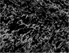
Samples were processed following routine histological procedures and stained using Verhoeff–Van Gieson (VVG) staining protocol for elastic and collagen fibers [14].
Digital images of VVG stained superficial fascia were acquired at 10× magnification using Olympus BX 43 microscope (Olympus America Inc., Center Valley, PA, USA). With VVG staining the elastic fibers appeared black while collagen fibers appeared pinkish in color. The images were analyzed using the TissueQuant software (SOIS, Manipal University, Manipal, India) especially designed for quantification of the color [1516]. This software has been validated and has been successively used in different studies [171819].
Biochemical study: hydroxyproline estimation
The tissues of the superficial fascia were dried at 60℃ for 24 hours and the constant dry weight was recorded. The dried tissues were then treated with 10 ml 6 N HCl and kept at 110℃ for 24 hours. The neutralized acid hydrolysates of the dry tissues were used for the determination of hydroxyproline content [6].
Statistical analysis was performed using the SPSS ver. 15 (SPSS Inc., Chicago, IL, USA). Data was expressed as mean±standard deviation and 95% confidence interval. Paired sample t test was applied for comparing upper abdomen and lower abdomen parameters in each sex. P<0.05 was considered statistically significant.
Results
Histological findings
The superficial fascia of abdomen was formed by loosely packed collagen fibers mixed with abundant elastic fibers and adipose tissue (Fig. 1).
Biochemical findings
Hydroxyproline content of superficial fascia of upper abdomen was significantly higher than in lower abdomen in both males (P=0.003) and females (P=0.003) (Table 4).
Discussion
The superficial fascia is formed of collagen fibers, loosely packed and mixed with abundant elastic fibers and adipose tissue (Fig. 1). Our findings support the previous studies [123].
The collagen fibers have great tensile strength. It can resist considerable tensile forces without significant increase in their length and at the same time they are pliable and can bend easily [12].
Elastic fibers run singly, branch and anastomose with other fibers. They are usually thinner than collagen fibers. Elastic fibers stretch easily with perfect recoil. These fibers are numerous in membranes that stretch periodically. Superficial fascia covering the anterior abdominal wall has high proportions of elastic fibers [12].
Our study reveals that elastic and collagen contents of superficial fascia are higher in upper abdomen than lower abdomen in both sexes. It was statistically significant in case of males, i.e., elastic contents (P=0.004) and collagen contents (P<0.001). This difference were however not significant in case of females. This may be due to fewer available samples (Tables 1, 2).
Hydroxyproline, an amino acid is found in collagen provides a direct measure of collagen content [67]. In the present study, hydroxyproline content was higher in upper abdomen than in lower abdomen, which further supports our histological findings of collagen content in upper and lower abdomen of superficial fascia (Tables 2, 3, 4).
Relatively lesser supporting elastic, collagen and hydroxyproline contents of superficial fascia of the lower abdomen may be the reason for the excessive bulges, excessive folds and relatively lower abdominal obesity. However, the estimation of the elastic, collagen and hydroxyproline contents of superficial fascia of the abdomen was not found in the available literature for comparison.
In abdominal obesity, fat cells lie between various layers of multilayered abdominal fascia [11]. Hence, for more favorable outcome, the study emphasizes that multilayered liposuction in different planes bound by multi layered abdominal fascia should be done. The knowledge of the elastic, collagen and hydroxyproline contents of the supporting abdominal fascia may help the surgeon in preoperative evaluation and to understand the outcome of liposuction in a given area.
Though the sample size was adequate, the number of female samples were less due to unavailability of female cadavers. A study including more number of female samples could provide more relevant results. The study gives a lead to further work on different age groups and comparison with this study and correlation between lean and obese individuals may be interesting.
Elastic, collagen and hydroxyproline contents of superficial fascia of upper abdomen were higher than lower abdomen. It is an initial observation on the basis of study of abdominal fascia. This may be one of the reasons for asymmetric bulging over abdomen and more sagging fold of skin in the lower abdomen than upper abdomen. The present study thus attempts to build a normal data base for the elastic, collagen and hydroxyproline contents of the superficial abdominal fascia.


 XML Download