

 PDF
PDF ePub
ePub Citation
 Print
Print


Introduction
Continuous attention has been developed on the anatomical variations of the axilla in anatomist and surgeon due to their clinical importance. One of the reasons is that it contains numerous neurovascular structures and lymph node continuing between the neck and the upper limb. Among the anatomical variations, axillary muscular variation may cause obstructions of vessels and nerves within the axilla region [1].
One of the most common muscular anatomical variation within the axillary region is Langer's axillary arch, which is called as variously axillopectoral muscle, pectodorsal muscle or arcus axillaris [234]. The axillary arch is generally in the form of a thin muscular slip and extends between the latissimus dorsi muscle and the pectoralis major muscle [5]. The prevalence of this variation muscle ranges from 7% to 27% [6]. It is clinically important surgeons to performing axillary surgery, especially breast reconstruction using the latissimus dorsi myocutaneous flap because of its close relationship to the neurovascular variations.
In present case, we report a case of the Langer's axillary arch accompanying neural variation, paying special attention to its clinical importance and embryological implication.
Case Report
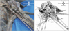
During the routine dissection of formalized cadavers for teaching students in Keimyung University School of Medicine, the upper limbs were dissected and observed carefully to study the compartments of the pectoral and axillary regions. Unexpectedly, in a 63-year-old male cadaver, we found variant muscular slips arising from the latissimus dorsi and inserting into the pectoralis major. As shown in Fig. 1, axillary arch muscle (also called Langer's mucle) was identified. This small muscular slip originated from the lateral border of tendinous part of the latissimus dorsi and continued 90 mm more crossing the axilla and lying superficial to the median nerve. This nerve was formed by the union of the lateral and medial cords of the brachial plexus, normally. After axillary arch crossed the median nerve, it inserted into the superior margin of the insertion of the pectoralis major. The variant was 92 mm in length and 7 mm wide.
Variation of the musculocutaneous nerve was also found in same side. The musculocutaneous nerve arose from the lateral cord of the brachial plexus, and it penetrated the coracobrachialis muscle. After that, it continued 107 mm more and connected to the median nerve. Other neurovascular structures showed typical patterns. On questioning the patient, there was no record about previous upper limb neurovascular symptoms.
Discussion
Axillary arch was mentioned firstly by Ramsay in 1795 and later it was confirmed by Langer in 1864 [1]. This variation was named as "axillopectoral arch," "Langer's muscle," "Langer's axillary arch," "arcus axillaris," "the pectodorsal muscle," or "axillopectoral muscle." Classically, axillary arch originated from the border of either latissimus dorsi or pectoralis major, whereas its insertions vary as following [7]. The latissimus dorsi-arising axillary arch muscle inserts into the fascia or tendon covering short head of biceps brachii, coracobrachialis, and pectoralis major. Some articles showed its insertion onto the medial epicondyle of the humerus and the medial intermuscular septum. Turgut et al. [8] reported a rare axillary arch originating from the coracoid process of the scapula and extending to the long head of triceps brachii muscle. In present case, axillary arch inserts into the pectoralis major, which is most common pattern by reviewing aforementioned cases. Interestingly, this small muscular slip crossed the median nerve perpendicularly and accompanied the variation of the musculocutaneous nerve. Previous cases also showed its co-incidence of the variations in adjacent nervous structures [910]. However, our case is extremely unique because of its deep association with the median and musculocutaneous nerves.
Axillary arch muscle is implicated in various clinical complications such as neurovascular compression syndrome including the entrapments of axillary vein, musculocutaneous, median, and ulnar nerves [1112]. Axillary arch muscle can be palpable during clinical examination and can be confused with enlarged lymph nodes and soft tissue tumors [13]. It is noteworthy to describe clinical significance of our case, which axillary arch crossed anterior to the median nerve perpendicularly.
According to Besana-Ciani and Greenall [11], the axillary arch muscle originates from panniculus carnosus, which is an embryological remnant of more extensive sheet of skin. Panniculus carnosus is well developed in lower mammals, while in higher primates and humans it is only evident as muscle such as platysma and dartos. In lower mammals, panniculus carnosus is highly developed to form pectoral group of muscle, however, in man it has regressed because its functional importance decreased during evolution in favor of wider upper limb mobility. Our and previous cases accompanying nervous variations suggested its embryological association with brachial plexus [910]. Its embryological and clinical significances should be investigated further with larger cases.
Here, we reported a rare axillary arch accompanying variation of the musculocutaneous nerve. Though most of axillary arch is usually asymptomatic, it may be associated with neurovascular structures accompanying clinical significance. Therefore, clinicians should be aware of this anatomical variation to avoid the misdiagnosis and complications during surgical interventions or procedure in axillary region.
 XML Download