

 PDF
PDF ePub
ePub Citation
 Print
Print


Introduction
The skeletal muscle injury of lower limb is one of the health problems that could reduce walking ability, so it can decrease the quality of life. Prevalence of muscle injury due to works or sports is about 30% of the population. Lower limb is part of the body that has a high risk of injury, because of the function in ambulation and bearing the body weight on the activities of daily living [12]. Women had the higher risk of muscle injury, due to the smaller sizes of their muscle mass, and the risk will be increased as they age [34].
The most frequent of the biomechanical abnormalities in the population was the functional leg length disparity, with a prevalence rate of between 60%–90%. There was an association between the functional leg length disparity with the pronated feet. The prevalence of the flexible pronated feet in the population was 20%–30% [56].
The formation of pronated feet was shifted of the talus bone to medial and caudal, causing internal rotation of the knee and the sacroiliac joints, tilted one side of the pelvic and created the functional leg length disparity [78910]. The series of biomechanical abnormalities will increase the eccentric contraction of the muscle, causing higher mechanical pressure of the muscles around the lower limb joints while doing walking activity, and initiated the skeletal muscle injury [1011]. The pronated foot formation usually occur whenever the heel strikes the ground on normal foot mechanism during walking activity, and will disappeared in the mid stance to start the supination activity, then followed by external rotation of the tibia and the sacro-iliac joints on the foot preparation to left the ground. Prolong pronated foot formation will disrupted the normal foot mechanism against gravity, thus increasing the tension on the lower limb muscles during walking activity [111213].
Walking activity is a repetitive progressive activity which the foot being a foundation for the leg. Individual who has biomechanical abnormalities experiences the repetitive muscle stress on the lower limb and increases the risk of muscle injury [131415]. Microstructure of the muscle cells will be changed after 5–15 minutes at the muscles activity [121314].
The skeletal muscle injury has suffered of the microstructure damage on the contractile elements, can caused leakaged of the creatine kinase-MM (CK-MM) enzyme into the blood plasma. The CK-MM enzyme was a protein found in the muscle cells cytoplasm, and served for converting the creatine ATP into phosphocreatine and the ADP. The CK-MM enzyme also play a role in the reformation of the creatine ATP [1516]. The CK-MM enzyme is an indicator of the microstructural damaged of the skeletal muscle tissue. The concentration will be increased in the blood plasma at 24–72 hours after the physical activity [171819].
It was necessary to correct the pronated feet, to prevent muscles injury and increase walking ability. The pronated feet correction by foot orthoses supporting the talus bone toward lateral and cranial, so center of gravity moved normaly to the second and third of metatarsals bone. Improvement of the foot bone formation expectedly to be improved of the knee and the hip joints formation during activity. Improvement of the biomechanical abnormalities will be reduced the risk of the muscle injury [20212223].
The flexible pronated feet needed a stable footwear to stabilize the structure of the foot during activity. The kind of footwear needed to support pronated feet is a solid and stable shoes. The solid and stable footwear prevents the foot joints from shifting abnormally during walk. The ideal footwear for pronated feet will cause the metatarsal area to be supinated while the heel leaves the ground (push off). The footwear should also had replaceable insole, which can be removed to accommodate the foot orthoses. The foot orthoses is placed inside the footwear as an insole providing the medial arc to support the talus bone [2223].
There is a wide range of the foot orthoses; from a simple prefabricated form that can be directly used as needed and easily found in the market, to the costumized form made according to the specific conditions of the patient's feet. There are some materials that are often used for the foot orthoses, i.e., the silicone, the ethylene vinyl acetate, and the polyethylene. The foot orthoses can be manufactured by making the foot print to duplicated any abnormalities on the feet, so can be designed the proper shapes of the foot orthoses [2223].
It was necessary to study the effect of the foot orthoses application as the correction of the pronated feet on muscle injury, by analyzing the CK-MM enzyme concentrations as the objective of muscle injury indicator in the blood plasma after walking test. The gender and the age of the subjects determined the rate of the muscle injury, thus to obtain the homogeneous subjects and have not experienced the degenerative problems, the study was done on the young women aged 20–29 years old [34].
It was discovered that the muscle injury occurs after highly intense of physical activity than the functional daily physical activity. For that reason, this study conducted the walking test to triggered the micro-muscle injury, by performing the maximal speed of the walking test as subjects could only tolerate 15 minutes. The preliminary study had to be done by several examination to determined when was the highest lever of the enzyme after a walking test [1314].
Materials and Methods
The study was a double-blind clinical trial with control. Research subjects were adult young women, who had biomechanical abnormalities of the pronated feet accompanied by the functional leg length disparity. Research subjects were divided into two groups, i.e., group that used the sport shoes within orthoses for pronated feet, and group that used the sport shoes without orthoses. The foot othoses used in this study was prefabricated polyethylene insole that can be remodeled by heating the material adjusted to the subjects foot form. The orthoses can easily found in the market.
Confirmation of the changes of talus bone as an effect of the foot orthoses, evaluated by X-ray radiograph have taken with subjects stood weight beared lateraly. The differences of the talus showed that talus bone was higher after using foot orthoses.
The CK-MM enzyme as the muscle injury indicator was assessed before and between 24–72 hours after the walking test.
Preliminary studies have been done on the randomized six subjects. It was conducted to determine the most appropriate time to get the highest level of CK-MM enzyme concentrations between 24–72 hours after the walking test. Evaluation of the CK-MM concentrations in preliminary study has done at 24, 40, and 45.
The study was conducted in West Jakarta, in October 2010 to February 2011. Sample size calculated based on the purpose of research was to analyzed the effect of using foot orthoses in the changed of CK-MM enzyme concentrations after the walking test by the two mean differences formula.
Based on calculation of the predicted sample size for each group, at least with the consideration of the probability sample drop out, it would require 14 subjects for each group.
Research subjects were young adult women ages 20–29 years old with abnormal biomechanical of pronated feet accompanied by functional leg length disparity. The inclusion criteria was healthy young women, willing to subject themselves to a series of studies that include the examination of the body, the blood, and walking test, as well as willing to sign an informed consent. The exclusion criteria was for patients who have history of lower limb trauma, lower limb pain, have neurologyc problem, which caused of walk disruption, have regular exercise three times per week on the last 2 weeks, and had another risk factor to have high concentration of CK-MM enzyme in blood plasma.
Their blood samples were taken from a median cubital vein. The blood samples were examined from clotted blood serum. Examination of the CK-MM concentrations by Hydragel 15 ISO-CK electrophoresis technique (Sebia, Norcross, GA, USA) was done between 24–72 hours after the 15-minute walking test.
The value of the CK-MM enzyme concentrations was obtained in the percentage of the total creatine kinase enzyme concentrations, so we need to access the concentration of total creatine kinase enzyme in the International Unit (IU). Examination of total creatine kinase concentrations was conducted by the kinetic method, 340 nm spectrophotometric.
Prior ethical approval was obtained from Universitas Indonesia Fakultas Kedokteran (No. 201/PT02.FK/ETIK/2010).
Results
Among the 177 young women aged 20–29 years selected, only 37 of the subjects (21%) met the inclusion criteria and did not had the exclusion criteria, 10 subjects were dropped out, and finally there were 27 subjects (15%) who were willing to follow the entire series of examinations in this study. Randomized allocation was performed using a computer program, 14 subjects entered the treatment group, and 13 subjects entered the control group. All the subjects were the university students at the west Jakarta.
Physical characteristics of the subjects
All the subjects have similarities of physical characteristics; the mean of age was 20.53±0.4 (P=0.33), the mean of body height was 1.62±0.4 m (P=0.58), the mean of body weight was 56.74±9.4 kg (P=0.83), and the mean of body mass index was 22.75±2.3 kg/m2 (P=0.42). The subjects also have similarities of demographics, activities and level of the education.
Preliminary study
The preliminary study had done on the six subjects selected by simple random sampling, shown that the highest level of the enzyme achieved at 45 hours after a walking test.
Intensity of the walking test
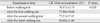
Intensity of the walking test was determined by the pulse frequency measurement (times per minute) of both group. Pulse frequencies before a walking on treatment group 86.43±13 was similar with control group (P=0.778), while pulse frequencies after a walking test on treatment group 146.86±11, as well as similar with control group (P=0.586), as indicated that both group had done relatively same intensity of a walking test.
Effect of the foot orthoses on the CK-MM concentrations 45 hours after the walking test
The examination of the CK-MM consentrations by the electrophoresis on hydragel showed that the 100% of the total creatine kinase enzyme on the subjects was the CK-MM enzyme. The CK-MM concentrations are shown in Table 1. The differences of elevation of the CK-MM concentrations before and after the walking test on both groups can be seen in the Table 2. The differences of the CK-MM enzyme concentrations before and 45 hours after the first, the second and the third walking test on the treatment group, which the interval of the each tests were 7 days, can be seen in Table 3.
The ANOVA repeated test analyzed the differences in the CK-MM concentrations and revealed the significant differences after those walking tests. The CK-MM concentrations after the first walking test was higher than before the walking test (P=0.007), and the CK-MM concentrations after the third walking test was lower than the second walking test (P=0.029). The resulted indicated there has physiologicaly adaptation on used the foot orthoses in the treatment group after the second walking test.
Discussion
The benefits of foot orthoses on the pronated feet correction is supported by several studies. Murley et al. [22] proved that there were the lower of the anterior tibial, the posterior tibial, and the peroneal muscle activities when the heel touched the ground by using foot orthoses [2425]. The study by Shorten [20] and Bishop et al. [21] also showed that there was an increase in the dorsiflexion motion, and reduction in the shiftd among the foot bones when the heel touched the ground while using the specific footwear within the foot orthoses inside the shoes [2326]. Nawoczenski et al. [27] proved that the benefits of the foot orthoses were the energy absorption, supported the medial arc, filled in the space between the foot and the shoe, and reduced the pressured on the foot area, so the leg muscles work lighter [24].
This study supported the previous studies that by using the foot orthoses, the foot could became supinated when left the ground. Foot orthoses supported the talus bone to the lateral and cranialy that has proved by the radiograph x-ray examination in this study. The tibia and the sacro-iliac joints could became externaly rotation. The improvement of biomechanical structure will cause reduce in the tension of intrinsic-extrinsic foot muscles, as well as improve the lower limb balance during walking activity. These improvement will reduce muscle injury.
We need to prove that pronated feet correction by using foot orthoses will decrease the risk of muscle injury by study of CK-MM enzyme concentrations as an indicator of skeletal muscle injury. This study found that concentration of CK-MM enzyme was higher in the control group which not using the foot orthoses. This study proved that the subjects have biomechanical abnormalities which not corrected by foot orthoses had higher degree of the muscle injury than the subjects which corrected by foot orthoses.
The elevation of the CK-MM indicates that there is an increase of number of the injured muscle. An injured muscle cells will be damaged in the sarcolema and cause the release of the CK-MM enzyme intracellular into the blood plasma [101115161718]. The studies done by Minkowsky and Minkowsky [28] and Weiner et al. [24] suggested that the series of the biomechanical abnormalities in the several joints, will increase the eccentric contractions and increase the pressure on the muscle tissues that worked on the abnormal joints during activity [101115]. The studies by Saxton et al. [25] and Eston et al. [29] found that high eccentric contractions on the muscle activity, will increase the risk of the muscle injury [2830].
The elevation of the CK-MM concentrations is lower in subjects which received the pronated feet correction. Concentrations of CK-MM enzyme continued to decrease after second and third walking test in treatment group. This result indicates that the pronated feet correction by foot orthoses will improve the series of biomechanical abnormalities, and will decrease eccentric contraction, reduce the pressure in muscle tissues, and finally reduce the number of muscle injury while walking. Decrease of CK-MM concentrations after using foot orthoses for several times, described that there was physiological adaptation in using foot orthoses, and the benefit of foot pronated correction will increase after long term using.
Limitation of this study were there is no data about the CK-MM consentrations between 45–72 hours after walking test.
Correcting of pronated feet on biomechanical abnormalities by foot orthoses in young women will reduce the degree of leg muscle injury. The reduce of muscle injury will be optimum after several times using of foot orthoses.
Recommendations
(1) It is necessary to use foot orthoses on young adult women who have biomechanics abnormality of pronated feet with functional leg length disparity, to prevent the occurrence of leg muscle injury during walking activity.
(2) Examination of the total creatine kinase enzyme before and 45 hours after physical activity can be done, as an objective indicator of the muscle injury degree in healthy young adult women.
(3) It is necessary to do the research to analyze the effects of foot orthoses in young adult males who have biomechanical abnormalities.
(4) It is necessary to do the research to analyze the concentrations of CK-MM enzyme 45–72 hours after physical activity.
(5) It is necessary to do the research to analyze the effects foot orthoses with different high medial arch support, and more economical foot orthoses materials, toward the degree of muscle injury indicator.
(6) It is necessary to do the research to analyze the effect of foot orthoses in young adults who have biomechanical abnormalities and also have the excessive body weight or obesity.


 XML Download