

 PDF
PDF ePub
ePub Citation
 Print
Print


Introduction
The uncinate process is a prolongation at the junction of the lower and left lateral border of the pancreatic head. The word "uncinate" comes from the latin "uncinatus," meaning "hooked" [1]. The pancreas develops from the two endodermal buds which arise from the caudal part of the foregut [2]. Most of the pancreas is derived from the larger dorsal pancreatic bud which appears first and lies cranial to the ventral bud. The smaller ventral pancreatic bud develops near the entry of the bile duct into the duodenum and grows between the layers of the ventral mesentery. After the rotation of the duodenum to the right side, the ventral bud lies posterior to the dorsal bud and later fuses with it. The dorsal pancreatic bud becomes the upper part of head, neck, body and tail of the pancreas and the ventral pancreatic bud forms the lower part of pancreatic head and uncinate process [3]. Malformations of the pancreatic uncinate process have been attributed to excessive fusion between the ventral and dorsal analogues during embryonic development [4]. A basic understanding of the embryologic development and normal anatomy of the pancreas and biliary tree is therefore essential for identifying these anomalies.
Numerous anatomical anomalies of the pancreas and the pancreatic ductal system are commonly encountered during radiological assessment. These pancreatic variants may simulate various neoplastic, inflammatory and post-traumatic conditions. Anatomical variations, developmental anomalies (e.g., pancreas divisum, annular pancreas, ectopic pancreas, pancreatic agenesis and hypoplasia) [5] and congenital diseases (congenital pancreatic cysts, von Hippel-Lindau disease, choledochal cysts) can all pose a diagnostic challenge for the clinicians [6]. Such anomalies should be borne in mind during differential diagnosis for abnormal conditions of pancreas and its associated structures. To the best of our knowledge such variant disposition of pancreatic head and uncinate process has not yet been reported during pancreaticoduodenectomy. It could be significant during surgical resection and pancreaticojejunostomy.
Case Report
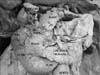
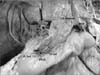
We encountered a rare variant in the constitution and disposition of pancreas in an adult male cadaver during the course of preclinical educational training program for undergraduate medical students. The pancreas displayed a usual J-shaped profile with unduly large uncinate process. The head of the pancreas was not confined to the concavity of duodenum. The enlarged uncinate process was found overlapping the third part of duodenum and a small peritoneal fold was observed connecting the inferior surface of the head of the pancreas to the left extremity of the third part of duodenum (pancreatico-duodenal fold) (Fig. 1). The head of the pancreas measured 4.5 cm in length and 6 cm in its maximum width. An unusual recess (pancreatico-duodenal recess) was formed bounded by the posterior surface of the enlarged uncinate process, anterior surface of the distal portion of third part of duodenum and the pancreatico-duodenal fold (Fig. 2). It measured 1.6 cm in length, 2 cm in breadth, and 2 cm in depth. On further exploration, the duodenopancreatic fold was found to be avascular. The enlarged head and uncinate process received vascular supply from inferior pancreatico-duodenal artery. The superior mesenteric vessels were related anteriorly to the uncinate process of pancreas. The pancreas displayed normal consistency and was not adherent to the underlying structures. The neck, body and tail of pancreas displayed normal anatomy.
Discussion
The pathological changes of the pancreas are occasionally related to its embryological development. Abnormal pancreatic developmental stages are associated with a variety of diseases of the gland. Pancreatic disorders can be classified as those affecting the ductal system and those related to the parenchyma of the gland [7]. Precise knowledge of the embryogenesis is necessary to the understanding and thus the treatment of many diseases of pancreas. One of the most interesting but rarely documented variations of the pancreas is hypertrophied uncinate process which may be due to excessive fusion between the ventral and dorsal buds during embryonic development [4]. The anomaly encountered in the current investigation could also be viewed as a consequence of deranged fusion of ventral and dorsal pancreatic buds. Sometimes the pancreas fails to develop normally and there may be congenital defects associated with the uncinate process. There are several instances of abnormal development of the pancreatic uncinate process, such as an annular pancreas, pancreas divisum [8]. Similarly, portal annular pancreas is another interesting anatomical anomaly in which the pancreatic tissue surrounds the superior mesenteric vein and portal vein like a ring but in a few cases the uncinate process of the pancreas has been found forming this ring [9]. These types of cases have to be considered carefully during pancreatic resections.
It is important for surgeons to acquaint with the anatomical relations of the uncinate process of the pancreas especially with the superior mesenteric vessels to avoid unwanted iatrogenic complications. The uncinate process, unlike the remainder of the organ, passes posterior to the superior mesenteric vessels [10]. In the present study, the hypertrophied pancreatic tissue was observed to overlap the third part of the duodenum and was directly related to the superior mesenteric vessels and abdominal aorta. This may be responsible for compression of the surrounding neurovascular structures. The etiology of such pancreatic hypertrophy is usually congenital but can seldom be acquired [2]. It may result in clinical complications like obstruction of the duodenum or pancreatitis. Additionally, as also seen in Annular Pancreas, complications such as obstructive jaundice, peptic ulcer, duodenal perforations and peritonitis may result [11].
The peritoneal fold encountered in the present investigation is an additional interesting observation. It connected the head of pancreas with the third part of duodenum and therefore, it is justifiably designated as pancreatico-duodenal fold. Further, the recess formed as a consequence can be referred to as pancreatico-duodenal recess. Understandably, it possibly can be an occult site for internal herniation and can cause diagnostic and intra-operative dilemma. Laparoscopic investigations are gaining popularity especially of the abdominal region. In the event of the presence of such congenital malformations it becomes imperative to acquaint oneself with possibility related to faulty embryogenesis. Further, this may avoid intra-operative complications.
In conclusion, endoscopists and surgeons should give utmost importance to such a rare pancreatic anomaly during diagnostic and therapeutic procedures. This report will be a significant addition to the present anatomical literature.
 XML Download