

 PDF
PDF ePub
ePub Citation
 Print
Print


Introduction
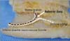
The mandibular canal containing the inferior alveolar neurovascular bundle crosses the mental foramen anteriorly and forms the anterior loop of the mandibular canal at the region where it splits into the mental and incisive canals [1]. The anterior loop continues beyond the anterior margin of the mental foramen, can be identified by the presence of the two separate canals [2], and develops during the double back process when the mental canal exits to the mental foramen from the mandibular canal [1].
At the same time the inferior alveolar neurovascular bundle passes through the mandibular canal, finally divides into two parts (the mental and incisive branches), and participates in the formation of the anterior loop. The mental branch supplies sensation to the skin and mucous membrane of the lower lip and chin together with adjacent buccal nerve, and the vestibular gingiva of the mandibular anterior teeth, while the incisive branch innervates the anterior teeth including the first premolar [3, 4, 5]. Therefore, a complete understanding of the anatomical structures in the interforaminal region containing the anterior loop is essential to prevent neurosensory disturbances resulting from direct or indirect damage to the neurovascular bundle during surgical procedures involving the mandible, such as dental implant installation, open reduction of a mandibular fracture, and genioplasty [6, 7, 8, 9].
Several studies have been conducted to identify the prevalence and location of the anterior loop, as well as anterior loop length (ALL). Previous researchers have set a plane that passes through the anterior-most margin of the anterior loop that is coincident with the origin of the incisive canal as a standard reference for the anterior loop [1, 6, 10]. A radiologic study set a cutoff point of 3 mm as the maximum diameter of the incisive canal as it separates from the anterior loop [2, 11]. However, standard references for the anterior loop differ according to the study method.
At the region where the inferior alveolar neurovascular bundle divides into the mental and incisive branches, the incisive nerve bundle is totally separated from the surrounding epineurium of the mental nerve bundle [12]. In the present study, a standard reference was defined for the anterior loop by locating the perineurium of the inferior alveolar neurovascular bundle, which divides into the mental and incisive nerve bundles, using the micro-dissection at the interforaminal region. The main aim of this study was to elucidate the general anatomical structure of the anterior loop of the mandibular canal in Korean cadavers using morphometry relative to the defined standard references.
Materials and Methods
The anterior loop of the mandibular canal was examined in 19 embalmed Korean cadavers (26 hemimandibles; 16 males and 3 females) with a mean age at death of 54.4 years (range, 29-75 years). These cadavers had been donated for educational purposes to the Department of Anatomy, School of Medicine, Chosun University. This study followed the Declaration of Helsinki with respect to the medical protocol and ethics.
All hemimandibles that had been taken to identify the intraosseous course where the mandibular canal ramifies were decalcified for 3 days in a decalcification solution (10% nitric acid), after which they were neutralized in distilled water for 12 hours. The buccal cortical and cancellous bone was then carefully removed, taking great care not to damage the inferior alveolar neurovascular bundle. The configuration of the mental foramen was also preserved with care. The mental and incisive neurovascular bundles were covered with a separate epineurium and had specific terminal distribution areas [12]. Bearing that in mind, the mandibular canal where the neurovascular perineurium divides into the mental and incisive branches were meticulously dissected with the aid of a surgical microscope (OPMI-FC, Carl Zeiss, Oberkochen, Germany).
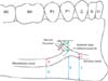
After determining the cutoff point of the neurovascular bundle, that point was determined as the anterior-most margin of anterior loop coincident with the origin of incisive canal (Fig. 1). Fiducial points were set based on this location defined as a reference point: the mandibular canal was 10 mm back from the reference point, and the incisive canal was 5 mm forward of the reference point. The diameters of the mandibular, mental, and incisive canal were measured at each reference point. Then, the distance was measured from the inferior border of the mandible to the inferior margin to the points 10 mm to the rear (i.e., mandibular canal), the reference point (the point that was ahead of the anterior loop and defined autonomously as stated above), and 5 mm forward of that point (i.e., incisive canal) (Fig. 2).
The topography of the anterior loop was investigated with reference to the mental foramen. Only the contour of mental foramen was preserved; the buccal alveolar bone was eliminated, which meant that it was possible for the location of the foramen to change. Therefore, the locations of the alveolar crest and the mental foramen by the inferior border of the mandible were measured before removal of the alveolar bone, and then had a conjugation for establishing the accurate landmark. The distance between the midpoint of the mental foramen and the anterior loop (i.e., the length of the mental canal), the horizontal distance between the anterior margin of the mental foramen and the anterior loop, and the vertical distance between the superior margin of the mental foramen and the anterior loop were measured (Fig. 2). Measurements were made using digital vernier calipers (Mitutoyo, Kawasaki, Japan) to an accuracy of 0.01 mm in all cases.
Two investigators measured the same items twice on subsequent days. Statistical analysis using one-way ANOVA was performed with SPSS version 12.0 (SPSS Inc., Chicago, IL, USA) to determine the mean, SD, interobserver difference, and difference between the left and right sides. The interobserver analysis indicated that there was no statistically significant difference between the values measured by the two investigators (P=0.847), and so the mean of each measurement pair was used as the final value of each measurement. There was no significant difference between the right and left sides (P=0.649). Furthermore, the diameter of each canal and the distance from the inferior border of the mandible at the reference point of each canal were evaluated using one-way ANOVA with a post-hoc comparison on Scheffé's method. No distinctions were made with regard to either age or gender. The data are presented as mean±SD values, and the cutoff for statistical significance was set at P<0.05.
Results
The diameters of the mandibular canal at the point 10 mm back from the anterior loop, at the mental canal, and at the incisive canal at the point of 5 mm forward of the anterior loop were 2.80, 2.63, and 2.22 mm, respectively. There was significant difference between the diameters of the mandibular and incisive canals. The distances from the inferior border of the mandible to the point 10 mm back from the anterior loop, to the anterior loop, and to the point 5 mm forward of the anterior loop were 7.82, 10.11, and 9.08 mm, respectively. The mandibular canal ascended to the area where the mental and incisive canals diverged, and then passed downward to the incisive canal. The distances between the inferior border of the mandible and mandibular canal and anterior loop differed significantly (Table 1).
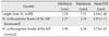
The distance from the midpoint of the mental foramen to the anterior loop was 4.34±1.46 mm. The mean horizontal distance from the anterior margin of the mental foramen to the anterior loop was 3.05 mm, and ranged from 1.17 to 5.18 mm; the distance was >4.0 mm in 19.2% of cases (n=5). The mean vertical distance from the superior margin of the mental foramen to the anterior loop was 2.72 mm, and ranged from 1.19 to 6.34 mm; the distance was >4.0 mm in 11.5% of cases (n=3) (Table 2).
Discussion
The anterior loop, which can be described as the extension of mandibular canal anterior to the mental foramen, is formed just before the ramification of the mandibular canal into the incisive canal [2]. It includes the mental and incisive nerves simultaneously; therefore, caution should be taken during surgical procedures in the interforaminal region to avoid nerve damage [2, 11, 13].
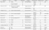
Many researchers have made attempts to detect the prevalence and location of the anterior loop, as well as the ALL in this context (Table 3) [1, 2, 6, 7, 14, 15, 16, 17, 18], and a safe guideline has been proposed for implant treatment planning in the distal aspect of the interforaminal area. However, the prevalence of the anterior loop has been reported as varying from 0% to 88% [11], and the recommended guideline varies greatly from 1 to 6 mm [8]. These wide ranges of the prevalence and recommended guideline may be attributable to interindividual anatomical variability associated with gender, age, and race, and the use of different measurement techniques [3, 8]. Especially on the measurement methods, the direct measurement using cadaver is very higher prevalence than the radiologic examination, and also cone beam computed tomography or spiral computed tomography providing higher resolution radiographs shows higher incidence of visualization to the panoramic radiograph [10, 15, 16, 17]. This may be explained that the identification of the anterior loop is affected by a calcification degree of the cortex which represents radiopaque in radiographic images [19].
In addition, it was thought that the difference in the standard used for defining the anterior loop was the most important factor. The present study micro-dissected the inferior alveolar nerve, which runs within the mandibular canal; the ramification point of each epineurium that covers the mental and incisive nerves was defined as the anterior loop, and the inferior alveolar nerve divided the mental and incisive nerves in all cases. However, because the shape of the anterior loop occasionally is a continuous straight pattern toward anterior teeth not forming the loop [12], further study is needed to determine the location of the ramification point according to the pattern of the anterior loop.
As with the ALL defined in previous research, in the present study the mean horizontal distance between the anterior margin of the mental foramen and the anterior loop was 3.05 mm. Although this anterior extension of the mandibular canal from the mental foramen shows a diversity of results with respect to side, age, and dental state in previous research, it was especially significantly longer for male, great height, and Asians race; thus, race-related physique is an important influencing factor [2, 10, 11, 15, 17, 18]. In addition, it is important to be aware of the longest measurement of the anterior loop in order to avoid nerve injury during surgery. Previous studies have found values of 9 mm in panoramic radiographs [20] and 11 mm in cadavers [17], whereas that in the present study was smaller at 5.18 mm. Additional studies must be conducted with larger samples to determine the true maximum value relative to gender, age, and race.
Full knowledge of the vertical distance below the mental foramen must be acquired to enable a safe sliding osteotomy during genioplasty [7]. In the present study, the average vertical distance below the superior margin of the mental foramen was 2.72 mm, with a maximum value of 6.34 mm. Hwang et al. [7] recommended a safety margin of at least 4.5 mm below the inferior margin of the mental foramen.
In the present study, the diameter of the mandibular canal decreased significantly from 2.80 mm at the 10 mm posterior point of the anterior loop, to an incisive canal diameter of 2.22 mm at the 5 mm anterior point of the anterior loop. On radiologic studies, the incisive canal can be identified in only 15% of images, with a reported diameter of 1.73 mm at the 4 mm anterior point of the anterior loop [20, 21]. Therefore, even though a radiologic examination should be performed as part of the treatment planning before surgical procedures pertaining to the incisive canal at the mandibular anterior teeth region, radiographs should not be solely relied upon for important morphological information, since such images suggest that the diameter is smaller than actual measurements made in cadavers, and moreover the visibility is poor.
In this study, the distances from the inferior border of the mandible to the mandibular canal, anterior loop, and incisive canal were 7.82, 10.11, and 9.08 mm, respectively, as they ran superiorly in order to exit the mental foramen, and then inferiorly and anteriorly to the chin after ramification. The findings that the mandible shape changes from a round shape in the posterior region to a buccal concavity in the anterior region, and that the vertical height of the mandibular body increases toward the anterior region must surely affect the actual location of each canal [7, 13]. However, the distance from the inferior border of the mandible to the mandibular canal at the level of the mental foramen remains constant throughout life [22]. Thus, the mandibular canal was located close to the alveolar crest at the anterior loop region where the ramification occurs, and the incisive canal was located higher than the mandibular canal from the inferior border of the mandible.
In conclusion, the anterior loop of the mandibular canal was located at a mean of 3.1 mm anterior and 2.7 mm inferior to the mental foramen, and continued upward and backward into the mental canal, and forward into the incisive canal. Since the mental and incisive nerves are each covered with an epineurium within the anterior loop, damage at the relevant region might cause dysesthesia not only at the anterior teeth but also at the lower lip and chin. The detailed morphological features of the anterior loop and related structures reported herein represent useful practical anatomical knowledge regarding the interforaminal region.

 XML Download