

 PDF
PDF ePub
ePub Citation
 Print
Print


Introduction
The major and minor salivary glands express nerve markers such as glial fibrillary acidic protein (GFAP; one of the intermediate filament proteins), S100 protein (a well known marker of glial and Schwann cells) and neuron-specific enolase (NSE) under normal conditions in the absence of cancer [1, 2, 3, 4]. These neural markers, as well as protein markers of smooth muscle (e.g., alpha smooth muscle actin; SMA) are usually used for classification of salivary gland tumors [5, 6, 7]. Using human midterm fetuses, Katori et al. [8] reported that, in contrast to the nearby submandibular gland, the sublingual gland has a neurogenic immunohistochemical character similar to that of small glands such as the palatal, pharyngeal and lingual glands.
Several studies using rats have indicated that a difference between the submandibular and sublingual glands is evident not only in the gland tissue itself but also in its innervation. Ekström et al. [9] described that, in contrast to the sublingual gland, the usual co-presence of the sensory nerve markers, calcitonin gene-related peptide and substance P, is absent in the rat submandibular gland. In a pharmacological study using rats, Mirfendereski et al. [10] reported a much stronger parasympathetic nerve response (to pituitary adenylate cyclase activating peptide) in the submandibular gland than the sublingual gland. Likewise, Takai et al. [11] reported a much denser supply of neuronal nitric oxide synthase (nNOS)-positive parasympathetic neuronal nitric in the submandibular gland than the sublingual gland of rats. Finally, also in rats, Tobin et al. [12] described that muscarinic receptor subtype M1 is predominant in the sublingual gland, in contrast to predominance of M5 in the submandibular gland, although M1 induces indirect effects via nitric oxide. These differences were not demonstrated in human specimens, possibly due to experimental difficulties.
Consequently, using specimens from elderly donated cadavers, the primary aim of this study was to comprehensively examine the immunohistochemical reactivity of usual marker proteins in the submandibular and sublingual glands. In addition to the aforementioned neural markers (GFAP, S100 protein, NSE, and nNOS), we also studied peripheral myelin protein 22 (PMP22), which has been used for staining of peripheral nerves (e.g., Gregson et al. [13]), and shows mutations in some forms of human neuropathy such as Charcot-Marie-Tooth disease [14]. Our secondary aim was to compare the expressions of these proteins between mucous and serous areas in mixed glands, for which we considered the best strategy would be to compare between mucous acini-predominant lobules with serous acini-predominant lobules.
Materials and Methods
The study was performed in accordance with the provisions of the Declaration of Helsinki 1995 (as revised in Edinburgh 2000). We examined 15 donated cadavers (7 men and 8 women) ranging in age from 78 to 95 years, with a mean age of 88 years. The cause of death had been ischemic heart failure or intracranial bleeding. These cadavers had been donated to Tokyo Dental College for research and education on human anatomy, and their use for research had been approved by the university ethics committee. The donated cadavers had been fixed by arterial perfusion of 10% v/v formalin solution and stored in 50% v/v ethanol solution for more than 3 months. From each of the cadaveric heads, we prepared 2 tissue blocks (the submandibular gland and sublingual glands). For easy discrimination of sections, the sublingual gland contained a small area of the oral mucosa. Thus, the sublingual gland was cut almost along the long axis, in contrast to the submandibular gland, which was cut transversely. After performing routine procedures for paraffin-embedded histology, 9-10 serial sections were prepared: two of them were stained with hematoxylin and eosin and with periodic acid Schiff reaction (PAS), respectively, while the others were used for immunohistochemistry (IHC).
The primary antibodies used for IHC were (1) rabbit polyclonal anti-human GFAP (1:100, Z0334, Dako Cytomation, Kyoto, Japan); (2) rabbit polyclonal anti-human S100 protein (1:100, N1573 or Z0311, Dako, Glostrup, Denmark); (3) mouse monoclonal anti-human NSE (1:100, N1557, Dako); (4) rabbit polyclonal anti-human PMP22 (1:1,000, p0078, Sigma-Aldrich, St. Louis, MO, USA); (5) rabbit polyclonal anti-human neuronal nNOS (1:200, Cell Signaling Technology, Beverly, MA, USA); (6) rabbit polyclonal anti-human tyrosine hydroxylase (1:500, ab152, Millipore-Chemicon, Temecula, CA, USA); and (7) mouse monoclonal anti-human alpha SMA (1:100, M0851, Dako). Among these antibodies, Nos. 1-6 were for demonstration of nerve supply to the glands, while No. 7 was for identification of myoepithelial cells. Except for the antibodies against S100 and SMA, antigen retrieval was performed using microwave treatment (500 W, 15 minutes, pH 6). The secondary antibody (incubation for 30 minutes, 1:1,000, Histofine Simple Stain Max-PO, Nichirei, Tokyo, Japan) was labeled with horseradish peroxidase (HRP), and antigen-antibody reactions were detected by HRP-catalyzed reaction with diaminobenzidine (incubation for 3-5 minutes; Histofine Simple Stain DAB, Nichirei). All samples were counterstained with hematoxylin. Samples without the primary antibody were used as negative controls. The present S100 antibody, N1573, reacts strongly with S100B protein but weakly or very weakly with S100A protein, whereas Z0311 is specific for S100A protein. The present anti-SMA antibody reacts strongly with the endothelium of arteries and veins [15]. We counted NSE-positive nerve elements in the whole sectional area of both mucous and serous lobules using ×10 or ×20 objectives. The data was evaluated using t test.
Results
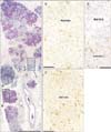
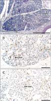
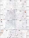
PAS staining clearly demonstrated a mosaic pattern formed by the mucous and serous acini in the sublingual and submandibular glands (Figs. 1, 2, 3). The sublingual gland was comprised of 30-60 round or triangular lobules in each section, while the submandibular gland had 60-120 quadrate, triangular or irregularly shaped lobules per section. Although the present observations were not three-dimensional, the lobules of the sublingual gland were classifiable into three patterns: (1) lobules composed purely of mucous acini; (2) lobules composed purely of serous acini; and (3) lobules containing both types of acini (i.e., actual mixed glands). The mucous lobules numbered 3-8 per section (around 10%), the serous lobules 5-15 (around 20%), and the others-accounting for the majority-were regarded as mixed lobules. However, in the submandibular gland, the mucous acini, if present, were restricted to a small area in each lobule. Thus, the majority of lobules were serous, and mixed lobules accounted for less than 10% of the total. In the interlobar tissue, nNOS-positive as well as TH-positive sympathetic nerves were easily found. However, we were unable to trace them to areas around the acini because of the limited nature of the present IHC (Fig. 3).
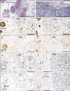
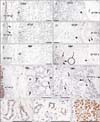
In both types of gland, myoepithelial cells expressed SMA, but IHC showed them to be negative for S100B (N1573 antibody), GFAP, and NSE (Figs. 4, 5). However, S100A antibody (Z0311) stained some of the myoepithelial cells and ductal cells in the sublingual gland (Fig. 3C). The positive part of the duct appeared to be intercalated, but most of the duct was negative. Thin nerve fibers in the arterial wall expressed GFAP (Figs. 4C, D, 5E, F). Blood cells as well as some of the submandibular ductal cells expressed PMP22 (Fig. 4F, I, J). The PMP22-positive part of the duct appeared to be the striated part, but most of the striated duct was PMP22-negative. In the interlobular tissue, thick myelinated nerve fibers expressed PMP (Fig. 4K) as well as S100 (Figs. 4A, B, 5C, D). Therefore, in the lobule, nerve elements along and around each of the acini were easily identified by IHC for NSE rather than for S100. SMA reactivity of myoepithelial cells was seen in both mucous and serous acini.
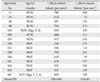
In the sublingual gland, the lobule containing serous acini showed a higher density of NSE-positive nerve elements than that containing mucous acini (Fig. 1B, C). The difference in density reached 10-fold at maximum: 300-800 NSE-positive cells per 1 mm2 in the serous lobule vs. 50-200 per 1 mm2 in the mucous lobule (Table 1). The difference in the sublingual gland was statistically significant (P<0.0001). Likewise, in the mixed lobule where serous and mucous acini were present in the same section, we found a clear demarcation between nerve-rich and nerve-poor areas (Fig. 1D). However, in the submandibular gland, where mucous acini were very sparse, the nerve elements were distributed almost evenly along and around the acini in any lobule (400-900 positive cells per 1 mm2). We examined and compared the NSE-positive nerve elements in the whole sectional area of both mucous and serous lobules using ×10 or ×20 objectives. Conversely, when we counted positive cells within a small area under a ×40 objective, limited nerve-rich areas were likely to be found even in lobules containing mucous acini. Finally, we did not find either a gender difference or a difference depending on ages.
Discussion
A well-known textbook [16] states that S100 expression is evident in the intercalated duct and myoepithelial cells of the major salivary glands. Although Katori et al. [8] expected that the adult sublingual gland would show strong neurogenic immunoreactivity of myoepithelial cells, the present study demonstrated negativity for S100B, GFAP, and NSE. In contrast, S100A antibody (Z0311) stained some of the myoepithelial cells and ductal cells. In the sublingual gland, the fetal neurogenic characteristics of myoepithelial cells, such as expression of S100 or GFAP, seemed to become reduced or disappear during postnatal life and/or with aging. Lee et al. [3] and Chi [17] stated that S100B reactivity was limited to fetuses, and was not present in adults. Likewise, Mori et al. [18] and Dardick et al. [19] reported that myoepithelial cells of the adult major salivary glands were negative for S100. As suggested by Korean pathologist [3, 17], the varying results for S100 immunoreactivity in references were probably due to poor discrimination between S100A and S100B by the antibodies employed. In the sublingual gland, S100B reactivity might disappear first during postnatal life, whereas S100A reactivity is likely to be retained during subsequent aging. We found PMP22-positive ducts in the submandibular gland. The positivity appeared to correspond to the striated part of the duct, but most of the duct was negative. Thus, PMP22 positivity may have reflected a specific physiological condition of the ducts, such as degeneration. Taken together, our findings suggested that a proportion of salivary gland cells retain neurogenic features during postnatal life.
To our knowledge, Kusakabe et al. [20] are the only group to have reported a difference in innervation between the mucous and serous acini in the human submandibular gland. In their excellent IHC study using cryostat sections of specimens from relatively younger patients, they found that neuropeptide Y-positive nerve fibers (candidate sympathetic nerves) as well as vasoactive intestinal polypeptide-positive nerve fibers (candidate parasympathetic nerves) were more densely distributed around mucous acini than around serous acini. They considered that such peptidergic nerves may play a more active role in regulating the secretory mechanisms in the former than in the latter. Although the difference we noted was not in the submandibular gland but the sublingual gland, the findings of Kusakabe et al. [20] differed from our present result. It seems difficult to evaluate differences in very thin nerves between adjacent small areas because irregular staining is liable to occur, especially in cryostat sections.
Careful evaluation of IHC results is necessary, using not only high-magnification but also lower-magnification views. In fact, when we counted NSE-positive cells within a small area under a ×40 objective, nerve-rich areas tended to be revealed even in lobules occupied by mucous acini. Therefore, it was not possible to effectively compare mucous and serous acini in the submandibular gland of elderly individuals because of the very small mucous area surrounded by a large serous area. In contrast, the sublingual gland of elderly individuals provided a good model for such a comparison because purely mucous lobules were present with purely serous lobules in each section. Using this model, we intend to further examine differences in nerve supply. The present counting of NSE-positive cells revealed a statistically significant difference between the serous and mucus lobules (Table 1). However, the positive cells were not always nerve-associated cells but they were likely to include neurogenic mesenchymal cells. The present method, counting cells, might not be the best way for a quantitative analysis of nerves.
 XML Download