

 PDF
PDF ePub
ePub Citation
 Print
Print


INTRODUCTION
The morphological and functional unit of the bone is the Haversian system, with textbooks diagrams showing Haversian or central (longitudinal) canals connected by Volkmann's or perforating (transverse) canals. Officially, Haversian and Volkmann's canals are "nutrient and perforating canal" in Terminologia Histologica [123]. The canals have a concentric lamellar organization and are of equal size. The bone is vascularized by vessels that penetrate the matrix from the periosteum.
The microstructure of the compact bone is important to bone remodeling and modification in pathologic conditions. Therefore, knowledge of the microstructure of compact bone such as the Haversian system is important to understanding remodeling activity, distribution analysis, the pathogenesis of the osteoporosis, and other mechanisms. However, many studies of the Haversian system have focused only on the canals themselves due to methodology limitations associated with cross-sectional histology and microcomputed tomography [4567], rather than on the general canal system or the spatial relationships between the two types of canals. Knowledge of the overall composition of the Haversian system as well as the constituent canals themselves is very important when investigating changes relative to the normal system.
The aim of the present study was to demonstrate the actual microstructure and morphologic pattern of the Haversian system of compact bone using histology and three-dimensional (3D) reconstruction, rather than relying on virtual images. This described reconstruction technique can provide a general view of the architecture of the Haversian system and further the understanding of pathologic progression of the compact bone.
MATERIALS AND METHODS
Materials and sampling
Twenty Sprague-Dawley rats aged 8-10 weeks were used. The animals were perfused with 150 ml of saline followed by 600 ml of fixative containing 4% paraformaldehyde in phosphate-buffered saline. The femur was harvested and decalcified in 5% EDTA-2Na. The specimens were trimmed to 3 mm to make them easier to handle and to facilitate the acquisition of images during fixation. Each specimen was obtained from the anterior midshaft of the femur.
Tissue processing
After dehydration and infiltration, the paraffin-embedded specimens were cut transversely into 150 to 200 serial sections at a thickness of 5 µm for 3D reconstruction. The sectioned tissues were then stained with hematoxylin and eosin.
3D reconstruction
All of the stained sections were photographed using a 2,048×1,536-pixel digital CCD camera (DP70, Olympus, Tokyo, Japan). The serial images were aligned manually using computer software, the Haversian and Volkmann's canals were segmented manually, and all structures were reconstructed three-dimensionally. "Reconstruct" software was used to produce the 3D images of the Haversian system (this software can be downloaded from http://synapses.clm.utexas.edu/tools/reconstruct/reconstruct.stm) [8].
RESULTS
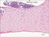
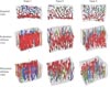
The Haversian canals were arranged into concentric rings and connected to each other by oblique perforating canals in the transverse sections (Fig. 1). However, the network pattern, spatial relationship between Haversian and Volkmann's canals, and difference between endosteal and periosteal sides were difficult to identify in the transverse histology sections. In contrast, all of these features were clearly evident in the reconstructed 3D models (Fig. 2). The basic structure comprises Haversian canals that are large in the endosteal region and highly interconnected. The canals in the endosteal region appear irregular and close to neighboring canals. In the other hand, the canals of the periosteal region are relatively small and less interconnected; that is, the network density is significantly higher in the endosteal region than in the periosteal region. The Haversian canals are straight and long and parallel to the length of femur. The Volkmann's canals run perpendicular to the Haversian canals, interconnecting the latter with each other and the periosteum, and extending in random directions at various angles.
DISCUSSION
Haversian canals are a series of tubes around narrow channels formed by lamellae. The Haversian canals surround blood vessels and nerve fibers throughout the bone and communicate with osteocytes. The canals and the surrounding lamellae are called a Haversian system (or an osteon). A Haversian canal generally contains one or two capillaries and nerve fibers. The spaces between Haversian systems contain interstitial lamellae. The osteonal pattern of compact bone is gradually built around the intracortical vessels by the progression of the cutting cones in a process of secondary remodeling; therefore, the central canal size can be used as an index of the remodeling activity [9].
Textbook diagrams of the Haversian and Volkmann's canals in the Haversian system indicate that the central canal size is equal in the endosteal and periosteal regions of compact bone [123]. However, we found that the Haversian system has a complex pattern of organization that is dominated by branching or Volkmann's canals. Our method of combining 3D reconstructions with histology examinations has confirmed the actual morphologic pattern of the Haversian system.
The main result of the present study is that the Haversian system is convoluted and closely interconnected in the endosteal region, while being straight in the periosteal region. The canals are significantly larger in the endosteal region than in the periosteal region. Moreover, there are more interstitial lamellae in the periosteal region.
The microstructure of the Haversian system is characterized by a branched network pattern that differs between the endosteal and periosteal sides. Because previous studies of the pathology of the compact bone during osteoporosis have generally focused on the thickness of the compact bone, the present results are useful in providing a general view of the architecture of the Haversian system and for understanding pathologic progression of the compact bone. In particular, in cases of osteoporosis the thickness of the cortical bone is reduced even though the external diameter remains almost intact. It might be that the degree of bone resorption is much greater in the endosteal region due to the presence of a large and highly interconnected Haversian system there.
The present results have demonstrated that the network of the Haversian system can be revealed only by combining 3D reconstruction with histology observations. We therefore further suggest that 3D reconstruction could also be a highly effective tool in other areas of morphology research.
 XML Download