

 PDF
PDF ePub
ePub Citation
 Print
Print


Introduction
Coordinated movements of eyeballs are important for binocular vision. These movements are brought by extraocular muscles. There are four recti muscles namely medial rectus, inferior rectus, lateral rectus, superior rectus, and two oblique muscles, i.e., inferior oblique, superior oblique [12]. An imaginary line joining the insertions of four recti forms a spiral pattern and is called spiral of Tillaxus. The precise location of insertions of these muscles is an important anatomical landmark while performing strabismus surgeries [3].
With advancement in diagnostic and treatment modalities, strabismus or squint surgeries have become more common. Apparent weakness or over action of any extraocular muscle on clinical examination is an indication of surgical intervention [4]. The aim of squint surgeries on extraocular muscles is to correct misalignment. Three types of surgical procedures are carried out in squint surgeries: (1) weakening-which decreases the pull of a muscle; (2) strengthening-which enhances the pull of a muscle; (3) procedures that changes the direction of muscle actions [3]. These procedures involve detachment and subsequent repositioning of insertion of muscles.
Materials and Methods
Forty eyeballs (20 right and 20 left) obtained from male adult embalmed cadavers from the collection of the Department of Anatomy were utilized for the study.
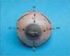
The cranial cavity was opened by cutting out the skull vault. After removal of brain the roof of the orbit was cut open to expose the contents. The neurovascular structures were cut near the apex of the orbit and the origins of extraocular muscles were also cut. Superior and inferior conjunctival fornices were incised to deliver the eyeball from the anterior aspect. The eyeballs were thoroughly cleaned and periocular connective tissue was removed. The insertions of the extraocular muscles were cleaned. Eyeballs were injected with the normal saline to maintain the integrity and shape of the eyeballs before taking the measurements (Fig. 1). This was required as it was observed that removal of eye balls from the orbit and cleaning of insertion involved handling of eyeball. Moreever cutting of vessels and nerves entering the eyeball left leakage points. The above two maneuvers resulted in leakage of fluid from within the eyeball causing distortion in shape and size and shrinkage. Following measurements were obtained:
- Distance of recti from limbus (sclerocorneal junction)
- Width of insertion of recti
- Length of aponeurosis of recti
- Distance between insertion of adjacent recti muscles
- Width of insertion of superior oblique and inferior oblique
- Minimum distance between insertions of superior oblique and inferior oblique
All the measurements were recorded by standard digital vernier caliper. The data obtained was tabulated and analyzed for descriptive statistics. Paired sample t test was applied to find the statistical difference between the right and left side. SPSS software (SPSS Inc., Chicago, IL, USA) was used for statistical analysis.
Results
Table 1 shows descriptive statistics of different study parameters of rectus muscles. It was observed that the distance of insertions from the limbus gradually increased from medial rectus to superior rectus and was quite variable (wide range).
The distance between the insertions also increased clock-wise from medial rectus to superior rectus. The aponeurosis of medial rectus was shortest and that of lateral rectus was longest.
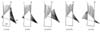
Table 2 shows descriptive statistics of insertion of oblique muscles. The superior oblique aponeurosis fanned out to be inserted beneath the superior rectus muscle. Fig. 2 shows various modes of insertion of superior oblique muscle with respect to superior rectus. Insertion of inferior oblique muscle was always fleshy. The muscle mostly inserted deep to the lateral rectus muscle except in six cases (4 right and 2 left) where part of insertion projected above the superior border of lateral rectus.
Discussion
Strabismus surgeries necessitate precise knowledge of insertions of extraocular muscles. Various surgical procedures like weakening or strengthening utilize the distance of insertion from the limbus as an important landmark.
The present study has documented the measurements of insertions of recti and oblique in Indian population.
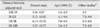
Findings of the present study on distance from the limbus are higher to the tune of 1 to 1.5 mm as compared to earlier studies (Table 3). There are no comparable findings in Indian population. The outcomes of the present study on width of insertions were about 1 to 1.5 mm less than earlier studies (Table 3). The distances between the insertions of adjacent recti were slightly higher than the earlier studies (Table 4).
Following points are noteworthy in regards to difference of findings of present study as compared to others:
(1) Most of the studies have been performed on white population.
(2) Precise methodology followed for measurement has not been described except by Apt [5]. He utilized eyeballs obtained from autopsy specimen that were fixed in formalin for five days followed by treatment with alcohol. However volume changes are bound to occur in any postmortem tissue, moreover shrinkage of tissue is known to occur after fixation in formalin and dehydration in alcohol. There is no mention of methodology in other studies. Observations by Sevel [6] are not comparable as they were recorded on fetal eyeballs.
(3) Besides racial distinction, authors wish to attribute this difference to the methodological dissimilarity. Authors observed that cleaning of the insertions of recti and obliqui and removal of the periorbital connective tissue required lot of handling of the eyeball. This resulted in leakage of fluid from the cut ends of neurovascular entry points in the eyeballs. This aspect was taken care in present study by perfusion of fluid, after cleaning the eyeball, for achieving the volume (and hence the size) of the eye ball as in living condition. Sclera is an indistensible tissue [7] hence possibility of stretching because of over perfusion is ruled out.
The length of aponeurosis of the lateral rectus muscle was longest and almost double the length of aponeurosis of other recti. The knowledge of lengths of aponeurosis will be of surgical importance as these are relatively avascular. Longer aponeurosis of lateral rectus may be due to longer lateral rectus muscle. Because of the obliquity of the eyeball, the distance of the insertion of lateral rectus from the origin of recti is longer and hence, longer aponeurosis. This fact has not received any mention in the available literature.
In a histological study of the lateral and medial recti, Jaggi et al. [8] have observed that there are no aponeurosis in the recti muscles and the muscle fibers are directly inserted on to the sclera. A naked eye examination however clearly indicates the insertion to be predominantly aponeurotic.
The aponeurosis of superior oblique was very variable in its position and extent. The range of width of its insertion was wide [8]. During eye surgeries identification of superior oblique is a tricky task. The position of superior oblique muscle with respect to the superior rectus can be a good guide for identification.
The findings of the present study may aid ophthalmologist during strabismus surgeries. The study also indicates towards need to reevaluate the insertion in vivo.


 XML Download