

 PDF
PDF ePub
ePub Citation
 Print
Print


Introduction
The coeliac trunk, the first ventral branch from the abdominal aorta, about 1.25 cm long passes almost horizontally forwards and slightly right above the pancreas and splenic vein, dividing into left gastric, splenic and common hepatic arteries.
Previous studies on cadavers and living individuals has shown manifold disparities of the coeliac trunk. About 15% of the population displays significant variations in the typical branching pattern [1].
The variations in the anatomy of the coeliac trunk must be carefully understood to enable anastomosing the appropriate arteries in postoperative closing. Additionally, knowledge about the variations of hepatic artery is important in liver transplantation, laparoscopic surgeries, partial hepatectomy, gastric resection, pancreatico-duodenectomy, radiological interventions and penetrating injuries to the abdomen. Furthermore the arterial variations are also important when matching organ procurements for transplantation.
Case Report
Variation in the coeliac trunk and its branches was observed during routine dissection of the abdominal region of a 60-year-old south Indian male cadaver in the Department of Anatomy, Kasturba Medical College (KMC), Manipal University, Manipal.
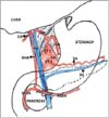
In the present case, a well-defined coeliac trunk was absent. The coeliac trunk was replaced by two separate arterial trunks at the level of lower border of T12 vertebra, 0.3 cm apart.
The first arterial trunk, about 2 cm long, after arising from the abdominal aorta bifurcated and provided the left gastric and the left hepatic arteries. Both the arteries when further traced had a normal course and termination. The left gastric artery reached the lesser curvature of the stomach to supply it. Few esophageal branches were also observed from the same. The left hepatic artery passed upwards to reach the porta-hepatis where it supplied the left lobe of the liver.
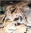
The second arterial trunk, about 1.5 cm long further bifurcated into a splenic artery and a hepato-gastroduodenal trunk. The splenic artery had a normal course and termination. However, the hepato-gastroduodenal trunk presented an unusual course. The artery moved to the right, further passing deep to the portal vein (Fig. 1). Here it provided the right gastroepiploic artery. The trunk then passed downwards and at the lower border of the neck of the pancreas, it emerged anteriorly. At this point, the artery bifurcated into right hepatic and superior pancreaticoduodenal arteries.
The right hepatic artery further ascended, passing anterior to the neck of the pancreas to reach the porta-hepatis to supply the right lobe of the liver. The cystic branch was provided by the right hepatic artery close to the porta-hepatis.
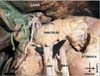
The superior pancreatico-duodenal artery when traced passed anterior to the head of the pancreas to reach the pancreatico-duodenal groove where it divided into anterior and posterior branches which further anastomosed with the corresponding divisions of the inferior pancreatico-duodenal artery arising from the superior mesenteric artery (Fig. 2). The superior and inferior mesenteric arteries when observed were normal. No other variations were observed. A schematic representation of the variations in the branching pattern of the coeliac trunk is provided (Fig. 3).
Discussion
The coeliac trunk is the first ventral branch of the abdominal aorta. In majority of the cases it arises as a single trunk (86%) and trifurcates to provide the classical branches, i.e., left gastric, splenic and common hepatic arteries [2]. However, two separate arterial twigs of the coeliac trunk as reported in the present case are rare.
Developmentally the sub-diaphragmatic ventral splanchnic arteries are paired initially and interconnected by the ventral and dorsal splanchnic anastomotic channels to supply the gut. Between sixth to tenth weeks of gestation, the mid gut undergoes rapid growth in a considerably smaller peritoneal cavity resulting in herniation, rotation and reposition. These changes cause hemodynamic alterations in the developing gut resulting in reduction of number of ventral splanchnic arteries into three as celiac trunk, superior and inferior mesenteric arteries. Alteration in either persistence or disappearance of these anastomotic channels and roots of these vessels may lead to variations in the celiaco-mesenteric origin and their branching patterns [34].
The left gastric artery, the smallest of the entire coeliac branches may originate directly from the abdominal aorta in about 1.9%-15% of the cases [25]. Variations of the hepatic artery are also well-described in the literature. The most commonly encountered variations are the aberrant hepatic arteries [1]. The aberrant hepatic arteries are further classified in to: the accessory and the replacing ones. A vessel that supplies a lobe in addition to its normal one is the accessory hepatic artery, while the replaced hepatic artery is the only branch provides the sole blood supply to that lobe, but originates from other than the normal position [4]. Most often an accessory or replaced right hepatic artery is seen to emerge from the superior mesenteric artery, while accessory or replaced left hepatic arteries seem to be branches of the left gastric artery [456789]. Other, possible locations of origin might be the gastroduodenal artery or the abdominal aorta [1489].
The left gastric artery emerging from the coeliac trunk and providing the left hepatic artery is reported and is assigned type II category in Michel's and Hiatt's classifications [1011]. However in the present case, a common trunk was observed which further bifurcated into left gastric and the replaced left hepatic arteries. The knowledge of these variations is important in intra-vascular procedures like arterial chemoembolization to prevent the spread of hepatic carcinoma and trans-arterial embolization to prevent blood loss in patients with severe hemoptysis [3].
The incidence of the bifurcation of coeliac trunk (common hepatic and splenic arteries) is not unusual and is reported [112]. However, our case had an unusual presentation, where in another common trunk arising from the abdominal aorta bifurcated in to: splenic and hepato-gastroduodenal trunk. Further the variant course and branching pattern of hepato-gastroduodenal trunk behind the pancreas and portal vein is an extremely rare occurrence and is not well documented and discussed in the literature.
Adachi [13] in his study mentioned, if and only if the hepatic artery emerges from the superior mesenteric artery (hepatomesentric trunk), then it may course behind the portal vein. Saga et al. [6] reported a similar case where the independent hepatomesentric trunk was coursing behind the portal vein. However, in the present case the anomalous hepato-gastroduodenal trunk was passing deep to the portal vein which then moved downwards and at the lower border of the neck of the pancreas; it emerged anteriorly to bifurcate into right hepatic and superior pancreatico-duodenal arteries. This type of presentation is rare and is seldom reported. The close association of these arteries to the portal vein may increase the risk of injury during resections of the pancreatic head [5]. Further, during laparoscopic surgeries, lack of awareness of these variations and limited operative field may lead to postoperative hemorrhage and ischemia or necrosis of the concerned organs.
Ramanadham et al. [14] reported a replaced right hepatic artery traveling anterior to the head of the pancreas just lateral to the neck of the pancreas. In addition, the gastroduodenal artery arose from the superior mesenteric artery caudal to the pancreas [14]. In the present case, the right hepatic artery was arising from the hepato-gastroduodenal trunk close to the lower border of the neck of the pancreas. It then ascended passing anterior to the neck of the pancreas to reach the porta-hepatis to supply the right lobe of the liver.
Normally, in all cases of abnormal origin of common hepatic artery, cystic artery begins close to the gall bladder in a twisted manner, which is more likely to get damaged in laparoscopic cholecystectomy [3].
The relevance of the hepatic vascular anatomy is of significance not only for harvesting and re-anastomosis of orthotopic liver transplants but also to avoid vessel injury in general surgery, particularly laparoscopic cholecystectomies and vascular radiological procedures. The abnormal course of the right hepatic artery also has a bearing on surgical and interventional radiological planning. In particular, since it is an end artery, its presence must be recognized when carrying out a pancreaticoduodenectomy procedure or dissecting out the portahepatis during resection of the liver and preservation of variant vessels during both right lobe liver living donor and split graft transplantation [15].
In conclusion, the complex variations in the coeliac trunk branches presented in our case are rare and highly significant as the coeliac trunk is the sole artery to the upper abdominal viscera. Preoperative knowledge of such rare variations is essential for clinicians to accomplish successful surgeries, diagnostic and therapeutic vascular intervention procedures and liver transplantations.
 XML Download