

 PDF
PDF ePub
ePub Citation
 Print
Print


Introduction
Dual left anterior interventricular coronary artery (also called left anterior descending coronary artery, hereafter referred as LAD), first classified by Spindola-Franco et al. [1], has been subdivided into six different types, based on the origin and course of the LAD [2]. In types I, II, and III, the LAD originates in the left coronary artery (LCA), and divides in two branches: the short LAD and the long LAD. In types IV, V and VI, the long LAD originates in the right coronary artery (RCA) or the right aortic sinus and the short in the LCA or the left aortic sinus.
Type IV dual LAD is an anomaly in which a short LAD originates from the LCA and a long LAD originates from the RCA. The long LAD passes anterior to the right ventricular outflow tract on its epicardial or intramyocardial course [12]. Type IV dual LAD accounts for 0.004% of dual LAD cases [3] and has never been reported in Koreans. We report a new variant of type IV dual LAD in a Korean cadaver.
Case Report
During a routine dissection carried out at Jeju National University Medical School in 2013, we found a case of type IV dual LAD originating from the LCA and RCA in a 57-year-old Korean male cadaver, whose cause of death was labeled "unknown."
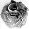
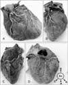
The LCA and RCA branched out of the left and right aortic sinus and had an external diameter (ED) of 4.9 mm and 4.4 mm, respectively (Fig. 1). The LCA was trifurcated into the LAD (ED 3.7 mm), the diagonal (ED 4.3 mm), and the circumflex (ED 3.8 mm) branches (Figs. 1, 2B). The LAD gave off a septal branch and coursed left to the anterior interventricular sulcus (AIVS) toward the apex cordis, the diagonal branch coursed left to the LAD toward the anterolateral wall of the left ventricle, and the circumflex branch coursed along the coronary sulcus. The other LAD (ED 1.7 mm) arose from the RCA 5.4 mm away from the right coronary orifice, crossed anteriorly to the infundibulum of the right ventricle, making a sharp turn just anterior to the pulmonary trunk to descend on the AIVS, but did not reach to the apex cordis (Figs. 1, 2A). The RCA gave off the posterior interventricular branch (ED 2.2 mm) which coursed along the posterior interventricular sulcus and terminated at the posterolateral wall of the left ventricle, where it met the LAD from the LCA (Fig. 2C, D).
Discussion
The frequency of dual LAD has been reported to be as low as 1% [1], but as high as 6.47% in the Republic of Korea [4]. Although type IV dual LAD is a very rare anomaly, it has been reported continuously [567]. The present report is the first case of a type IV dual LAD in a Korean cadaver based on the classification system, meaning a LAD by a septal branch to the AIVS [8]. One LAD originated from the RCA and traveled in the AIVS, which was the same as in the previously described long LAD [12], but it did not reach the apex cordis in this case. The other LAD arose from the LCA, never entered the AIVS through its course, but reached the apex cordis, where it met the posterior interventricular branch of the RCA.
These results met the classification of type IV dual LAD, but also had subtle differences. First, the LAD from the RCA did not continue to the distal AIVS in this case. It could have been considered as a short LAD, rather than a long LAD, which would not satisfy the standard of dual LAD. In type VI dual LAD, however, the long LAD from the RCA continues to the mid or distal AIVS [29]. Therefore, the LAD from the RCA could be considered to be a long LAD and then the dual LAD in this report might be classified as a new variant of type IV dual LAD, irrespective of the long LAD's continuation to the distal AIVS.
Second, the diagonal branch was one of the trifurcated branches from the LCA, not a branch of LAD, while the septal branch originated from both LADs, which has not been reported to date. The septal and diagonal branches have been described as major branches from both short and long LADs [12]. The septal and diagonal branches are known to originate from the short LAD. However, in type IV dual LAD, they are known to originate from the short LAD, not from the long LAD. Diagonal branches arising from both LADs have only been reported in type VI dual LAD [29], but its significance has garnered little attention. Similarly, our findings on the septal and diagonal branches might need further study to become clinically relevant.
Finally, the LAD and the diagonal branch from the LCA can be partially considered as the real LAD. It is evident that the artery originating in the RCA travels only halfway to the subinfundibular area, winds back toward the right side, and does not travel throughout the AIVS. Thus, the artery might appear as a right ventricular branch rather than one of dual LADs, which might be the limitation of the present case. This anatomical variation might be considered as a "third coronary artery" [10]. We, however, considered it as a new variant of type IV dual LAD since (1) each LAD originated from the LCA and the RCA, (2) each LAD is has a septal branch to the AIVS, and (3) the LAD from the RCA coursed as a typical long LAD, except it continued to the distal AIVS.
A LAD arising from the RCA is most commonly associated with a congenital coronary anomaly, tetralogy of Fallot (4%) [11], which generally follows the right ventricular outflow tract until it reaches the AIVS [12]. The high prevalence might represent the vestiges of the developmental position of the coronary orifices. The left coronary orifice is more posterior in tetralogy of Fallot than in normal hearts [1314], which might be related to an aortopulmonary rotation [15]. The dextroposition of the aorta could explain the more anterior position of the right coronary orifice and potentially the abnormal branching of the LAD to the RCA, as it crosses the subpulmonary infundibulum [14]. Therefore, abnormal positioning, especially more anterior positioning, of the right coronary orifice should be of concern whenever a variant coronary artery, such as a dual LAD, is on the right ventricle.
In conclusion, this is the first report on a new variant of type IV dual LAD in Koreans to the best of our knowledge. Although type IV dual LAD is not necessarily associated with unfavorable events, clinicians, as well as anatomists, should be aware of this variation to avoid misinterpretation of coronary artery distribution and further complications of clinical procedures.
 XML Download