

 PDF
PDF ePub
ePub Citation
 Print
Print


Introduction
The enteric nervous system (ENS) is considered the second brain in humans [1]. It is a constituent of the peripheral nervous system, which is distinctive because of its independence as it enables the intestine to manifest reflex activity in the absence of central nervous system inputs [2]. The ENS comprises a vast number of neuronal and glial cells equivalent to that of the spinal cord, which controls essential functions such as motility, secretion and blood flow [13]. Regardless of its high degree of complexity and its similarities to the central nervous system, the ENS is derived from the neural crest cells (NCC) which is the source of all of the peripheral nervous system [4]. It is derived from the somatic NCC at somite level 1-7 and then from 28 onwards [567].
The non-neuronal cells which appear before 10 weeks of gestation (WG) are amongst the first differentiated cells in the human gut wall and provide the first morphological elements of the ganglionic microenvironment [8]. The neuroblasts starts migrating from the neuroepithelium in about 5WG and reach the presumptive rectum in 7.5 to 12WG [9]. The submucosal plexus forms approximately 2-3 weeks after the myenteric plexus (MP) and arises from cells that migrate centripetally through the circular muscle layer from the MP [10]. By 14WG, foetal gut gave a mature appearance [11]. The peristalsis of the human foetal small intestine has been recorded from 12WG [12] but the oroanal peristalsis begins only around 27-30WG [8] and swallowing commences at 16WG [13]. The morphological and neurochemical properties of the human ENS are different from other species, even during development, therefore an in depth investigation of human ENS development is necessary. The study of the development of the ENS needs to be undertaken in the foetal gut as the ENS of the human gut is relatively mature at birth [214]. Development of the ENS in various segments of gut (like oesophagus, small intestine etc.) has been studied in details, but the literature is scant on the development of MP in the sigmoid colon of humans, which is a site of various congenital and acquired diseases. The development and maturation of the ENS in the sigmoid colon is important as many emergency surgical procedure conducted on it. The present study was designed to evaluate the maturation of the human sigmoid colon during various gestational time periods.
Materials and Methods
The human foetuses (n=12) were used from the foetal repository in the department of anatomy, All India Institute of Medical Sciences (collected at different times from the labour room of department of obstetrics and gynaecology, All India Institute of Medical Sciences). All the human foetuses were collected according to the protocol approved by the Ethics Committee, All India Institute of Medical Sciences, New Delhi. The foetuses less than 20WG were taken from cases where medical termination of pregnancy was performed for family planning (legalized in India under MTP Act, 1971) while those more than 20WG were stillbirths. All the mothers had no medical illness during pregnancy and none of the foetuses had any congenital anomalies. The foetuses were weighed and measured for crown rump length, foot length and biparietal diameter [1516]. The foetal age was determined according to above parameters and clinical history. The foetuses were immersed in 4% buffered paraformaldehyde after making a paramedian incision in the abdomen for proper fixation. Most of the foetuses used in the present study were obtained within 6-8 hours after delivery and were preserved at 4℃ to minimize any post-mortem changes. The human foetal sigmoid colon was identified by its mesentry and dissected out immediately.
NADPH diaphorase histochemistry-an enzyme histochemistry technique was used to study the tissue preparations. The colonic tissue was fixed in fresh 4% buffered paraformaldehyde for 2 hours at 4℃. They were washed thoroughly after fixation in chilled 0.1 M phosphate buffer. Cryoprotection was done by 15% and 30% sucrose for 3 hours and 8 hours at 4℃ respectively. The samples were then frozen in optimum cutting temperature compound (Sakura Finetek, Torrance, CA, USA) and 20-µm thick sections were cut using a Leica cryostat (Leica Microsystems Nussloch, Heidelberger, Germany). Frozen sections were mounted on 1% gelatin coated slides. Slides were kept at -20℃ for enzyme histochemistry. Cryostat sections on the glass slides were washed several times with 0.1 M phosphate buffer (pH 7.4). NADPH diaphorase activity was visible by incubating the sections in 10 mL 0.1 M Tris-Cl buffer (pH 7.8, adjusted with few microlitre of concentrated HCl) containing 10 mg NADPH (Sigma, St. Louis, MO, USA), 1 mg nitrobluetetrazolium (NBT) and 0.3% Triton X-100 at 37℃ for 45 minutes to 1 hour in the incubator in dark. The treatment with Triton X-100 is required because it is a good permeabilizing agent. The reaction was monitored under a dissecting microscope and were terminated by washing the tissues gently with chilled 0.1 M phosphate buffer when the stain was sufficiently intense (45 minutes-60 minutes). The sections were mounted in a mixture of glycerol and phosphate buffer (4:1) [17].
The stained sections were examined under a microscope and images were captured using a charge-coupled-device camera connected to a frame grabber card in an IBM PC interfaced with a Zeiss binocular microscope (Oberkochen, Germany). The images were saved in JPEG format with minimum compression and maximum quality. The images were then analysed using ImageJ (developed at the US National Institute of health, available at http://rsb.info.nih.gov/ij/). Before making measurements the system was calibrated using a micrometer scale (Carl Zeiss) for the magnification at which the images were acquired. More than 900 cell profiles were studied. The parameters used for analysis were as follows:
- Neuronal cell profiles (area, perimeter, and feret diameter).
- Myenteric fraction was calculated by the ganglionic area divided by area of muscularis externa in a section [13].
- Numerical density was calculated as by the total number of neuronal cells in the ganglionic area divided by area of ganglion multiplied by 7 µm.
- Number of neuronal cells per ganglion (the counting was done manually).
The SPSS software package version 17.0 (SPSS Inc., Chicago, IL, USA) was used for statistical analysis with the help of the Biostatistics Department of the All India Institute of Medical Sciences. The data was expressed as mean±standard deviation (SD). The data was analyzed by using Mann-Whitney test which is a non-parametric test. It was used to determine the statistical significance between the means of the computed data from various fetus. For all statistical tests, probability levels of less than, or equal to 0.05 (two-tailed P<0.05) were considered to be significant. According to gestational age, foetuses were assigned into two groups (group 1 [n=7], less than <17WG and group 2 [n=5], more than >17WG) for quantitative assessment.
Results
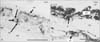
The NADPH-diaphorase-positive neuronal cells and their processes were identified in the MP of the sigmoid colon. A distinct pale nucleus was visible with surrounding dark blue cytoplasm in the neuronal cells. The neuronal processes and neuropil were also stained deep blue. The neuronal cells were packed inside the myenteric ganglia. The non-neuronal cells such as endothelial, mast, smooth muscle, glial cells and mucosal epitheliocytes did not take up the stain.
Group 1 (n=7)
At 14WG, the MP appeared as a continuous band of small neuronal cells. Binucleate neuronal cells were often observed. Large oval to round nuclei were there with little cytoplasm characterized the neuronal cells. Neuronal processes were observed emerging from the surface of the soma. Among the neuronal cells only simple neuropil was present at this age. Very few nerve fibers ran parallel to the circular muscle fibers. Scattered neuronal cells were present in the submucosa towards inner border of the circular muscle (Fig. 1A). At 15WG and 16WG, continuous band of cells formed into the elongated ganglia. Ganglia and neuronal cells were larger in comparison to the previous age group. There was obvious increase in the neuropil also. The nerve fibers were seen to colonize inside the circular muscle which were parallel to long axis of muscle fibers. Submucosal plexus was noted close to the circular muscle layer (Fig. 1B).
Group 2 (n=5)
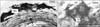
At 18WG to 20WG, ganglia were well organized. Neuronal cells were apparently bigger and the neuropil could be clearly visualized. The neuronal cells were pleomorphic with oval to rounded nuclei. Nerve fibers in the circular muscle were increased in number with increase in their thickness. At 23WG, the myenteric ganglia were increased in size. There was remarkable increase in the neuropil and nerve fibers in the circular muscle. There was an increase in the thickness of the nerve fibers also. In the submucosal plexus, large neuronal cells with thick fibers were noted (Fig. 2A). Large irregular neuronal cells with oval nuclei and thick and processes were observed (Fig. 2B).
Morphometric evaluation
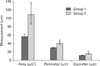
The morphometric data of the nitrergic neuronal cell profiles in both the groups were compared. It included the following parameters: mean area, perimeter, and feret diameter of the diaphorase positive neuronal cells. The average area of neuronal cells in group 1 was 57.31±4.66 µm2 and in group 2 it was 109.85±23.14 µm2 respectively. The perimeter of the neuronal cells in group 1 was 30.31±1.43 µm, and in the group 2 it was 41±3.73 µm respectively. The maximum diameter or feret diameter in group 1 was 11.81±0.62 µm and in group 2 it was 15.80±1.30 µm. Thus, all the parameters which were taken to assess the overall size of the neuronal cell in the MP were significantly increased (P=0.024) (Table 1, Fig. 3).
The mean of myenteric fraction in group 1 was 0.30±0.03, whereas in group 2 it was 0.32±0.02. The myenteric fraction was increased in group 2 but it was not statistically significant (P=0.33) (Table 1). The mean of numerical density in group 1 was (68.83±29.86)×105/mm3 and in group 2, it was (61±15.87)×105/mm3. The numerical density decreased in group 2 but it was not statistically significant (P=0.71) (Table 1). The mean number of neuronal cells per ganglion in group 1 was 11.89±4.33 and in group 2, it was 13.50±5.60. The number of neuronal cells per ganglion was increased in group 2 but it was not statistically significant (P=0.71) (Table 1).
Discussion
To our knowledge, this study quantified for the first time, some of the morphological changes in the innervation of foetal sigmoid colon during second trimester in Indian population. In this study, the maturation of the neuronal cells was assessed by analyzing the neuronal cell profiles in MP which has increased from group 1 to group 2. There was gradual increase in the neuronal cell size from 14-23WG which was statistically significant in NADPH-diaphorase stained neuronal cells (P=0.024). This increase in size may be due to increase in the cytoplasm as the size seems to be related to cellular maturity indicating higher rate of protein synthesis with increasing age. Hitchcock et al. [13] observed that nerve cell size increased with maturation from 6 µm at 8WG to 20 mm at term and 21 µm at 28 months postnatally in the MP of human oesophagus. These authors have shown that neuronal cell size increases greatly during fetal development and grows very slowly after its function is established. This size increase is associated with an increase in cytoplasm and the onset of peptide expression [13]. Gabella has also documented there is increase in the neuronal cell size with age in neonates and ageing guinea pig ileum [19].
In our study, numerical density of cells was less in group 2 in comparison to group 1 and especially from 20 to 23WG, but it was not statistically significant (P=0.71). This needs further investigation with the number of samples increased and using modern stereological morphometric methods. According to one study on the oesophagus, the numbers of cells, nerve density and myenteric fraction all peaked at 16-20WG and then density decreased towards adult levels, the myenteric fraction decreased during the late second trimester and became constant from 30WG [13]. A similar trend is noted in our study although it was not statistically significant which can be resolved by studying the total number of neuronal cells in the MP using advanced morphometric methods (using stereology). This may be due to growth of the bowel with increasing surface area, reduction in the cell number due to apoptosis, or increased neuropil in the developing enteric nervous system. It is known that during the growth of the neuronal cells, the number of dendrites and axons also increases, thus these processes push the adjacent neuronal cells, so the numerical density may change with maturation in the MP [20]. Montedonico et al. [21] also studied the nitrergic numerical density in the submucosal plexus of the porcine distal bowel from fetal life to adulthood and observed a similar trend.
The myenteric fraction was increased in group 2 in comparison to group 1, but it was not statistically significant (P=0.33). Hitchcock et al. [13] studied neuronal cells with their nerve fibres within the MP and quantified them together as the myenteric fraction, similar to the present study. This fraction was similar to the cell count, that was maximal at 16-20WG and decreased during the late second trimester, but became constant from 30WG [13]. This also needs further confirmation by increasing the sample size and using advanced morphometric methods. Similarly, the number of neuronal cells per ganglion increased in group 2 in comparison to group 1 but it was not statistically significant (P=0.71). Authors claimed previously that the number of cells per ganglion also decreased in late second trimester and this may be due to actual reduction in the cell number [13].
This study presents a detailed quantitative analysis of the development of MP in human sigmoid colon during second trimester. During prenatal life there is an increase in the neuronal cell size from 14-23WG signifying maturational process. The results reveal that this maturational event increase after 17WG and extensive innervation is established at 23WG. The limitation of the present study was reduced sample size due to non-availability of foetuses and autolysis of tissue. Such studies are important to distinguish the normal and pathologic development of the ENS and hence it may help in linking the theory and clinical practice.

 XML Download