

 PDF
PDF ePub
ePub Citation
 Print
Print


Introduction
The lateral cord of the brachial plexus gives its first branch, the lateral pectoral nerve (LPN) which pierces clavipectoral fascia and enters deep surface of pectoralis major muscle to supply it. The remaining part of the lateral cord divides into musculocutaneous nerve (MCN) and lateral root (LR) of median nerve (MN). The MCN pierces coracobrachialis (CB) muscle and then passes obliquely down to the lateral side of the arm between biceps brachii (BB) and brachialis muscle. In its course through the arm it supplies CB, BB, and brachialis muscle. The branch to CB arises from MCN close to its origin before it pierces CB. The branches to BB and brachialis are given off after MCN has pierced CB [1].
The medial pectoral nerve (MPN) branches from the medial cord, supplies and pierces pectoralis minor to enter pectoralis major.
The ulnar nerve also arises from the medial cord (C8 and T1) but often receives C7 fibres via a communicating branch from the LR of MN.
The MN is usually formed anterior or anterolateral to the third part of axillary artery by the union of a medial root (MR) (from medial cord) and a LR (from lateral cord) [1].
Variations in the origin, formation and branching pattern of brachial plexus, its relations with axillary artery and absence of or communication between its branches are com mon [2, 3, 4]. But the simultaneous presence of multiple variations is a rare occurrence. The present investigation strives to report a unilateral occurrence of multiple variations in branches of medial and lateral cord of brachial plexus along with intercordal neural communication between these cords.
The presence of these variations makes the nerves vulnerable during routine neural repair and other surgical interventions in the pectoral and axillary region.
Case Report
During routine dissection for medical undergraduate teaching programme, standard skin incisions were made to expose the region of the right axilla and arm of a 58-year-old male cadaver. The pectoralis major and minor muscles were reflected laterally after detaching them from their origins, to expose the axillary neurovascular structures.
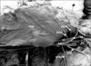
The lateral cord was giving off 2 LPNs-superior lateral pectoral nerve (SLPN) and inferior lateral pectoral nerve (ILPN) (Fig. 1). SLPN was seen to pierce the clavipectoral fascia and enter the pectoralis major muscle from its deep surface to supply it (comparable to a usual LPN). The ILPN was seen to arise from the lateral cord 1 mm distal to the origin of its superior counterpart and was observed to pierce the pectoralis minor muscle 5 mm inferior to its superior border.
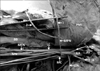
The MPN was given off from the medial cord of brachial plexus as usual. The length of the main trunk was 3 cm and it divided into two branches-a cranial branch (branch 1) entering the lower part of pectoralis minor and a caudal branch (branch 2) which was seen to supply pectoralis major without piercing pectoralis minor (Fig. 2).
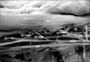
The MCN branched as usual from the lateral cord of brachial plexus but it did not pierce CB muscle (Fig. 3). Instead, it descended medial to the CB giving 4 branches (b1, b2, b3, and b4) and further pursued a normal course placed between biceps and brachialis. The first branch, b1, which was a short transverse branch to CB, was given off 2 cm distal to the origin of MCN and 4 cm from the coracoid process. It was seen to split into two before supplying CB. One centimeter distal to the origin of b1, a long oblique branch (b2) was given again to CB which disappeared within its substance to reappear again, posterior to the branch b3 and rejoining the main trunk of MCN caudal to b3. A third branch (b3) was given to BB, 6.2 cm caudal to origin of b2. The fourth branch (b4) was given to BB and brachialis as a common trunk at the same point where b2 rejoined the main trunk.
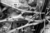
The MN was formed as usual by the union of MR and LR of MN arising from the medial and lateral cords of brachial plexus respectively. The MR of MN crossed the 3rd part of axillary artery from medial to lateral side to unite with the LR of MN which was 1 cm in length. In addition two neural communications were observed connecting the lateral and medial cords (Fig. 4). The cranial communication (* in Fig. 4) was arising from the lateral cord 3 cm proximal to the origin of LR of MN and the caudal communication (# in Fig. 4) was arising 1 mm distal to the cranial intercordal neural communication. Both the communications were seen running obliquely downwards and medially, anterior to the axillary artery and joining the medial cord. The cranial communication was bifurcating into two branches before joining the medial cord.
Furthermore, ulnar nerve was seen to arise by two roots from the medial cord-root "a" and root "b" (Fig. 4). Root "b," 1 cm in length, arose at the level of intercordal neural communication and root "a," 2.5 cm in length, arose higher up in relation to the 2nd part of axillary artery. Both these roots joined to form the main trunk of the ulnar nerve.
The arterial pattern and the relations of axillary artery to the nerves were found to be normal. No such variation was observed in the left brachial plexus.
Discussion
Variations in the nerves supplying upper limb have been reported by many authors [2, 3, 4, 5, 6, 7]. But multiple neuroanatomical variations coexisting in the same case involving the medial as well as the lateral cords have not been reported often in the literature.
In the present case, both the pectoral nerves showed deviation from the normal pattern. There were 2 LPNs, one supplying pectoralis major and the other supplying pectoralis minor. The MPN divided into two branches, one supplying pecto ralis major without piercing pectoralis minor and the other supplying pectoralis minor only. One similar case has been reported in which 3 pectoral nerves were seen to supply pectoralis major muscle only [2]. The MPN and LPN arose as a common trunk from the middle trunk of brachial plexus [3] in another study. Origin of LPN as 2 separate branches from the anterior divisions of upper and middle trunks instead of lateral cord is also documented [4]. The anatomy and course of these nerves is very important during breast surgeries using pectoral muscle flaps and pectoral nerve transfers to innervate paralysed arm in traction injuries of brachial plexus [5, 8].
The MCN normally pierces medial aspect of CB muscle, approximately 5 cm distal to the tip of coracoid process. Durgesh and Rao [6] reported the MCN descending parallel to medial border of CB without piercing it at any point. They found that branch to CB was arising directly from lateral cord [6]. In another study the MCN after piercing CB has been found to join the MN [7]. Rarely, lateral cord of brachial plexus may pierce CB and then divide into MCN and LR of MN [9]. Mavishettar and Iddalagave [10] reported that intercommunication between MCN and MN is 17.5%. However in this case there was no communication between MCN and MN. In shoulder joint traumatology, flap dissections, explorative procedures, post-traumatic evaluation of the arm and in performing axillary block or stimulating peripheral nerve in practice of anaesthesia this nerve and its course assumes great importance. The intrafascicular distance of each branch of MCN is important in microsurgical procedures [10].
The presence of intercordal neural communications bet ween medial and lateral cord of brachial plexus are uncommon [11]. Surgeons dealing with neoplasm or trauma repair need to be aware of these variations when they handle cases involving axillary artery and brachial plexus cords. The close course of the unusual intercordal communications, with the axillary artery, may compress the vessel and affect the blood supply of upper limb.
The uniqueness of the current case anomaly is also attributed to the origin of ulnar nerve by a second root at the level of intercordal neural communication between lateral and medial cords. This observation suggests the possible contribution of lateral cord as well in the origin of ulnar nerve.
Significant variations in the nerve patterns may be a result of altered signalling between mesenchymal cells and neuronal growth cones or circulatory factors at the time of fusion of brachial plexus cords [12]. Once formed, any developmental differences persist postnatally.
Communication between the medial and lateral cords of brachial pexus and origin of ulnar nerve by a second root at the level of this intercordal communication can be considered as a remnant from the phylogenetic or comparative point of view. Miller [13] summarized the work which was done by earlier authors on the brachial plexus in amphibians, reptiles, birds, monotremes, dogs, lemurs, primates in general and in anthropoid apes. He observed that only two cords-anterior and posterior, are seen in birds because the birds possess very little real forearm and hand structures. The ulnar nerve and MN are undifferentiated and a single nerve trunk supplies the region in the wings of birds. With progressively greater development in shoulder musculature and forearm and hand muscles, formation of 3 cords-medial and lateral cords anteriorly and a single cord posteriorly, has been observed in marsupials, lemurs and monkeys. The anterior cords however undergo fusion to give rise to MN and ulnar nerve. Thus, separation of nerves appears more definitely with increase in development of hand and finer muscle co-ordination. The complete separation of medial and lateral cords with ulnar nerve being a product of medial cord only is characteristically evident in man.
The nerves to the pectorals, the anterior thoracic nerves are three in number in species like birds, lemurs and monkeys, in which pectoral sheet is divided into major, minor and abdominal elements [13]. In the context that ontogeny recapitulates phylogeny; it is possible that the variations seen in current study is the result of a developmental anomaly.
The axillary region is very commonly approached by anaesthetists, orthopedicians, radiologists, and plastic surgeons. Lack of knowledge of these neural variations of brachial plexus may lead to intraoperative and post operative complications resulting in sensory, motor and trophic signs and symptoms. Additionally, adequate knowledge of neural and vascular variations may prove helpful in interpreting various clinical signs and symptoms with precision.
 XML Download