

 PDF
PDF ePub
ePub Citation
 Print
Print


Introduction
Volvulus is a condition of axial rotation of a part of the bowel through its mesentery [1]. The parts commonly involved in the volvulus formation include the caecal and sigmoid regions of the colon [2]. The factors such as presence of mesocolon for ascending or descending colon, very long colon are predisposing factors for a volvulus formation. Small bowel volvulus is a relatively rare cause of intestinal obstruction and could be fatal [3]. We report a rare case of congenitally displaced small and large bowels with a high risk of developing a volvulus at any stage of life.
Case Report
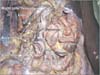
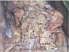
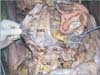
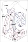
During dissection classes for medical undergraduates, we observed some congenital anomalies of the gut in an adult male cadaver aged approximately 70 years. The ascending colon ascended straight up to the right lobe of the liver. It gradually inclined to the left after reaching the right lobe of the liver to attain a midline position, where it suddenly turned down to continue as the descending colon. Thus there was a total absence of transverse colon. Terminal part of the ascending colon and the descending colon had a persistent mesocolon (Figs. 1, 2). Lower end of the descending mesocolon continued with the sigmoid mesocolon. Only the sigmoid mesocolon and the lower part of the descending mesocolon were attached to the posterior abdominal wall. Upper part of the descending mesocolon had a posterior free border containing the left colic artery. The terminal ileum passed to the right, deep to this free border to end in the caecum (Fig. 3). The descending colon was situated immediately to the left of the ascending colon throughout its course. The sigmoid colon began on the right side of the midline and coursed to the left to reach the left iliac fossa. It had a normal course into the pelvis thereafter. The jejunum and ileum were situated in the upper left part of the abdomen. They had a short mesentery. The ascending colon, descending colon and sigmoid colon were longer than usual due to the total absence of the transverse colon. A simplified illustration of the anomaly is given as Fig. 4. There were no other observable anomalies of the viscera and vessels of the abdomen.
Discussion
Most of the congenital anomalies of the bowel are dependent on the rotation of the midgut during its development. Midgut develops in 3 stages. In the first stage, midgut loop rotates through an angle of 90° in an anticlockwise manner as it enters the umbilical cord. In the second stage, the intestinal loop returns back into the abdomen from physiological hernia, at the end of tenth week of intrauterine life. At this stage, the caecum lies in the subhepatic region. In third stage, caecum and appendix descend caudally from subhepatic region, pass through right lumbar region and finally reach the definitive position in right iliac fossa. The total range of rotation around superior mesenteric artery is about 270°. After the completion of the rotation, the derivatives of midgut undergo a process of fixation [4]. If the fixation of the intestine is defective, parts such as ascending colon and descending colon will retain their mesentery. In the current case, the descending colon has failed to fall to its left and lose its mesentery during its development. This could be the possible reason for the absence of transverse colon and abnormal position of the small intestine also. In our literature survey, we could not come across any case of total absence of transverse colon and we assume that this is the first report on the absence of transverse colon. In the current case, mesentery of the small intestine was very small and the entire ileum and jejunum occupied the upper left part of the abdomen. One of the predisposing factors for the small intestine is the presence of a long mesentery [5]. The short mesentery and the congested condition of the small intestine in the abdomen might also lead to its abnormal movements leading to the formation of a volvulus. There are very few reported cases of persistent descending mesocolon and the volvulus of descending colon [6]. However, recently a few cases of right sided descending and sigmoid colons [7] and sessile ileum and subhepatic caecum [8] have been reported. In the current case, since the descending mesocolon was continuous with the sigmoid mesocolon, the chances of formation of volvulus are high. Since there was a gap behind the upper part of the descending mesocolon, the ileum of jejunum might pass through it and get strangulated at the gap.
To the best of our knowledge, total absence of transverse colon has not been reported yet. Absence of the transverse colon, presence of a descending mesocolon and displacement of small and large intestines as reported here might lead to one or the other complication at any time of life. The knowledge of this variation is very useful for the radiologist, gastroenterologists and surgeons in general.
 XML Download