

 PDF
PDF ePub
ePub Citation
 Print
Print


Introduction
Knowledge of vascular variations is of clinical and surgical importance. Indeed knowledge about the vascular variations as an essential pre-requisite in relevant invasive interventional diagnostic procedure could reduce possible morbidity and mortality [1]. Most of the vascular variations are asymptomatic and usually detected during the surgical procedures such as angiography or morbid anatomy studies. Among the vascular variations, arterial ones have been reported in various forms, but multiple absences of the branches of main arteries accompanied with other developmental abnormalities is an extremely rare variation. The Abdominal aorta (AA) is the largest and main artery in abdominal cavity. Classically its pattern of branching has been described as paired and single branches. Celiac trunk (CT), superior mesenteric artery (SMA), and inferior mesenteric artery (IMA) provide oxygenated blood to embryonic gut and its derivatives, while the paired visceral branches of AA supply diaphragm and retroperitoneal glands including kidney and ovary [2]. The pattern of branching of AA exhibits diverse variations both in level and types of branching. Various publications have reported diverse and multiple variations of the pattern of branching of AA including different arising levels of arterial branches, separated branches of CT, combined or fused arterial trunks, absence of one branch and arterial duplication [3]. In spite of a vast number of reported cases on variations of the branches of AA, absence of its multiple branches accompanied with other vascular, heart and endocrine abnormalities have not been reported. Obviously, such rare variations are of clinical importance particularly in interventional procedures and abdominal surgeries. Here we are reporting a very rare type of multiple absences of the branches of AA with congenital absence of the portal vein (CAPV), congenital unilateral adrenal agenesis and persistent ductus arteriosus in an adult female cadaver.
Case Report
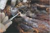
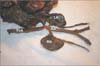
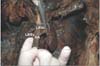
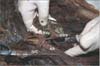
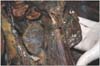
During a routine educational dissection of an adult unidentified woman who was dead due to car accident (30 years old; weight, 45 kg), and after complete dissection and opening the anterior wall of abdomen, the right free border of lesser omentum, hepatoduodenal ligament, was exposed and dissected to find the portal triad. Further inspection revealed that CT and its branches were absent. Therefore, the peritoneum of posterior abdominal wall was removed carefully. Following arterial variations were observed: (I) instead of CT, SMA, and IMA, solely a single arterial trunk aroused from (II) the anterior aspect of AA at T3-T4 level that (III) entered and distributed in mesentery (Fig. 1). Further inspection revealed that (IV) inferior phrenic and (V) ovarian arteries are absent in both sides while renal arteries showed no variation (Figs. 2, 3). Interestingly right and left ovarian veins exhibited ordinary course and drained to inferior vena cava and left renal vein respectively. After complete arterial exposing, we inspected kidneys and found (VI) two veins drained left kidney as follow; anterior renal vein run horizontally to right side in front of aorta and other behind AA and drained to inferior vena cava (Fig. 4). (VII) There were not superior, splenic and mesenteric veins, while left renal vein received an additional vein, which run downward and drained primarily all parts of digestive tract and its associated gland (portal vein did not exist) (Fig. 5). After meticulous inspection of renal vessels, we examined both kidneys and noticed that (VIII) right adrenal gland was absent (unilateral agenesis). Other findings included two pair's small lumbar arteries. Due to the extensive arterial variations of the branches of AA, we suspected to possible heart anomalies, so after opening the chest wall we removed and dissected the heart of cadaver. An interesting finding was persistent ductus arteriosus (PDA) (Fig. 6).
Discussion
We presented here the most uncommon multiple arterial variations accompanied with CAPV, unilateral right congenital adrenal gland agenesis (UCAA) and persistent ductus arteriosus which have not been reported before. The stomach, liver, spleen, pancreas, small and large intestines were supplied by a single artery which stemmed from the anterior aspect of AA at level of T3-T4. Additionally the right and left ovarian arteries were absent, while their venous pattern showed normal course. Interestingly the portal vein was absent and venous return of digestive tract were drained by a single vein which itself ended in the left renal vein (CAPV). Actually, these extensive vascular variations (multiple asence of the arterial branches of AA and CAPV) with heart anomaly and UCAA suggest a cardiovascular/endocrine syndrome which has not been reported. The embryological basis for such multiple variations can be explained as follow. Each primitive dorsal aorta gives off ventral splanchnic arteries (to the embryonic guts), lateral splanchnic (to the mesonephric ridge) and somatic arteries (supply the body wall). CT, SMA, and IMA are derived from embryonic ventral splanchnic arteries and provide blood supply to three primitive embryonic guts. The ventral splanchnic branches undergo a series of developmental changes including migration (descending), fusion and regression. In case of CT, in addition to descending from cervical region to subdiapharagmatic position, three separate branches (left gastric, splenic and common hepatic) are united by a series of anastomoses and finally CT is formed. The lateral splanchnic arteries including suprarenal, testicular and ovarian arteries persist on each side and retain their position [4]. In our presented case, the predominant embryonic scenario has probably been "regression." In case of absence ovarian arteries, the only reasonable explanation may be that after ovaries descending to pelvic cavity, their arteries (the lateral splanchnic arteries) regressed. In tandem with arterial scenario, venous system develops during embryonic period. Portal vein is formed by vetilline venous loop around the duodenum of the digestive tract, and then enters the septum transversum during 5-10 embryonic weeks [5]. With respect to the embryological description, it seems the CAPV in our presented case was secondary to the arterial variations and the portal vein was replaced with a vein which was drained to left renal vein. CAPV is an extremely rare variation (abnormality) and there are few reports on such abnormalities. Venkat-Raman et al. [6] reported CAPV in a fetus. Another case of CAPV was reported by Northrup et al. [7]. Based on previous studies (totally reported 18 cases) there are 2 subtypes of CAPV. In type Ia, the superior mesenteric vein (SMV) and the splenic vein (SV) do not join; therefore, no portal vein is formed anatomically. In type Ib, the SMV and SV do join in a normal manner, but this confluence drains to the IVC, not the liver. Type Ia CAPV is associated with atrial septal defect, ventricular sepal defect, and PDA [7]. Given the findings in our study, we propose a third subtype a single vein unit left renal vein with PDA and UCAA. Another set of our findings were multiple arterial variations. Based on the examination of abdominal organs (liver, spleen, small intestine and kidney; unpublished data) in one hand and multiple absence of the branches of AA in the other hand, we prefer to call these arterial variation as arterial anomalies. One of these anomalies was the absence of ovarian arteries. Although there are many reports on the variations of ovarian arteries [8], bilateral absent of ovarian arteries is a rare variation and only in one case bilateral absent of ovarian arteries has been reported [9]. With respect to the extensive absence of the arterial branches of AA, our reported case could be considered as one of the most uncommon arterial variation has been reported so far, because most of the previous literatures have reported cases with variations of the pattern of branching of AA including variations in origin of branches, level of branching, unusual course, common origin of arteries and single absent of one of the visceral branches of AA [3]. Among the reported variations, CT variations has been reported and studied extensively. According to previous reports CT shows a wide range of variations which have been analyzed and classified in detail based on the patterns of branching. For instance, Lipshutz [10] described different types of CT variations including hepatosplenic, gastrosplenic and celiacomesenteric trunk, but absent CT was not described. Complete absent of CT is a rare variation and its prevalence has been estimated between 0 to maximum 2% [11]. Morita [12] put forwarded a classification method of CT variation. According to Morita [12] four types of CT has been describes as follow as: 1) CT (textbook type), 2) hepatosplenic trunk, 3) gastrosplenic trunk, 4) hepatogastric trunk, and 5) absent CT [12]. Yi et al. [13] reported a rare variation of the absent of the CT in a Japanese cadaver, with the left gastric, splenic, common hepatic, and superior mesenteric arteries branching independently from the AA. Matusz et al. [14] described a case with absent of the CT, while the left gastric artery, common hepatic artery, and splenic artery originated directly and independently from the AA. Wu et al. [15] reported a 69-year-old female complete absence of SMA and compensatory dilation of the IMA. Accordingly he proposed a new classification method of superior-inferior mesenteric arterial variation (SIMAV). In this method SIMAV has been divided into four types as follow: type I, the normal type; type II, absent SMA; type III, absent IMA; and type IV, there is an aberrant middle mesenteric artery [15]. As we have seen in the present case, many visceral branches of AA were absent and additionally absence of portal system with other anomalies recommend a new revision in classification of such variations. The combination of multiple variation of arterial branches of AA with CAPV, PDA, and UCAA presented in our study suggest a new syndrome in living bodies. To the best of our knowledge it is the only reported case with such widespread anomalies. We think the importance of such case is beyond the surgical consideration and needs more profound developmental studies.

 XML Download