

 PDF
PDF ePub
ePub Citation
 Print
Print


Introduction
Ureteric stones are a common cause of obstruction of the urinary tract [1]. Typically, the patient has severe, intermittent pain that radiates downward from the loin into the groin as the stone travels from the kidneys down the ureter and into the bladder. The severity of the pain can be extremely intense, due to the spasm of the smooth muscle in the wall of the ureter, and it is often referred to the back, flank or groin regions, depending on the location of the stone and the level of obstruction [2, 3, 4]. Complications of ureteric stones include hydronephrosis, renal damage and infection of the urinary tract [2, 3]. Occasionally, stones may present with non-specific symptoms such as lower back pain and remain unidentified, resulting in growth of the stone and leading to chronic ureteric obstruction and complications, including hydronephrosis and renal damage. Hydronephrosis is a condition arising from obstruction to the urinary tract, causing dilatation of the ureter and calyces due to accumulation of urine. If mismanaged or untreated, hydronephrosis can result in permanent kidney damage and eventually may lead to renal failure [3]. The frequency of ureteric stones in patients with non-specific symptoms (silent stone) was recently reported as 0.7% [5]. There is a limited number of reports in the literature on hydronephrosis observed in cadavers. We report a case of a huge ureteric stone lodged at the left ureterovesical junction, associated with ipsilateral kidney shrinkage noted in a formalin embalmed old male cadaver, and discuss its clinical significance.
Case Report
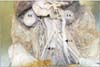
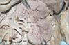
We observed a huge ureteric stone lodged at the left ureterovesical junction of an old male cadaver. The stone completely obstructed the ureter, as the saline injected into the proximal part of the ureter could not pass through the obstruction into the urinary bladder. The left ureter was grossly dilated with an average diameter of 15 mm compared to the right one (5 mm) throughout its entire length (200 mm). Compared to the right kidney, left ureteral wall was approximately 1.5-2 mm thick. Compared to the right kidney, left kidney appeared shrunken with presence of a nodular surface. The vertical length between the upper and lower poles of right and left kidney was 110 mm and 60 mm respectively. In addition, the width (between the hilum to the lateral border) of the right and left kidney were 50 mm and 40 mm respectively (Fig. 1). No other abnormality was detected proximal to the observed obstruction. Dissection of the distal part of the left ureter revealed a huge stone (measuring 25 mm×11 mm) encircled with a mass of white debris (Fig. 2). The dark-coloured hard stone which was removed, weighed 2.90 g and measured 1.6 ml in volume in a scale container. The stone seemed to be homogeneous in colour with a rough solid surface. In sagittal section, the left kidney appeared to have a dilated pelvis, as well as distended calyces filled with soft brownish post-mortem extravasations (Fig. 3). The right and left renal arteries were located behind the renal veins. The right and left renal veins were 20 mm and 45 mm in length respectively, with approximately similar diameters of 12 mm. The right and left renal arteries were 35 mm and 25 mm long respectively. The diameters of the right and left renal arteries were 7 mm and 8 mm respectively.
Discussion
Urinary stone formation is a long chronic process, which may be associated with a mild to severe symptoms for the patients [6, 7]. Obstruction of the ureter can give rise to increased ureteric and renal pelvic pressure resulting in severe hydronephrosis, with reduced glomerular filtration rate, tubular function, and blood flow of the kidney [1]. In addition, the ability of tubules to transport important electrolytes (such as Na+ and K+), and to adjust the urine concentration may be severely impaired [3, 6]. The severity of hydronephrosis is associated with the duration and extent of the obstruction [3]. Partial ureteric obstruction or brief disruptions may be limited to reversible functional disturbance, and may allow full recovery of renal function when corrected.
However, chronic obstruction of the ureter might lead to progressive renal damage and possibly tubular atrophy and permanent nephron loss, resulting in irreversible loss of renal function [3, 6]. In fact, back pressure of urine can reduce the blood flow of the kidney causing ischemic atrophy and urine extravasation, which may damage the renal tissue and consequently resulting in interstitial fibrosis, leading to kidney atrophy [8, 9]. Hence, this could explain why we observed a shrunken left kidney associated with a large ureteric stone and gross dilation of the ureter on the same side.
Specific ureteric stone-related symptoms, include acute ureteric colic, acute or chronic flank, abdominal or groin pain, hematuria, dysuria, anuria, or urinary tract infection [1, 10]. Colicky pain due to the presence of ureteric stones is well-known, but occasionally may be masked by the concurrent presence of other non-specific symptoms, such as backache. Zulkifli et al. described the case of a 43-year-old male, whose persistent backache masked the presence of his ureteric stone for more than two years, because the clinician believed the origin of the backache to be musculoskeletal, and no X-ray was taken throughout this period [11]. The pain from ureteric colic is often so severe that afferent pain impulses spread within the central nervous system, giving rise to nausea. Other possible symptoms of ureteric stones are vomiting, fever, lethargy, headache, hypertension and tachycardia [4, 7]. Patients with ureteric stones almost always present with hematuria, with approximately 90% of patients experiencing gross or microscopic hematuria. However, hematuria may rarely be absent, and this may indicate complete ureteric obstruction [1].
The symptoms of ureteric stones may also be non-specific in spinal cord injury (SCI) patients. Vaidyanathan et al. conducted a retrospective review of four cases of hydronephrosis due to urinary stones in SCI patients, in whom their diagnoses and treatment were delayed because these patients did not manifest the classical signs and symptoms of urinary stones. These non-specific symptoms comprise of feeling generally unwell, spasms, autonomic dysreflexia and abdominal discomfort. They concluded that the symptoms of urinary stones and hydronephrosis can be bizarre (non-specific) in SCI patients [12].
Ureteric stones can present with abdominal, back, flank or groin pain [3]. These symptoms may be similar to those of common mechanical lesions of the lumbar spine and pelvis, such as sacroiliac joint syndrome [13]. Acute ureteric colic may mimic the symptoms of acute abdomen or pelvic disorder [1]. In certain situations, the mechanical back pain of musculoskeletal origin and underlying pain arising from ureteric stones, may be similar in clinical presentation. Mechanical pain may appear together with the visceral pain of ureteric stones, thereby, confusing the attending clinician. Patients may exhibit non-specific soft tissue tenderness and/or muscle spasms in the lower thoracic or upper lumbar regions [4]. In less severe cases, a musculoskeletal diagnosis may be given if an incomplete patient history is taken. Practitioners with a specific focus on the musculoskeletal system, thus need to be aware of alternate causes of abdominal, back, flank and groin pain that are non-musculoskeletal in origin, such as ureteric stones [3]. Potential pain of visceral origin must be explored in cases of apparent somatic pain, since pain of visceral origin may be perceived as the latter, resulting in a potential overlap of symptoms of visceral and somatic conditions [13]. Other atypical symptoms of ureteric stones include bilateral inguinal pain, periumbilical pain, posterior hip pain and penile paresthesia [4, 13].
In conclusion, for a patient who presents with non-specific symptoms such as lower back pain, an accurate and thorough patient history, detailed physical examination and relevant investigations such as a kidney ureter bladder X-ray, ultrasound, or a computed tomography scan, could be helpful to rule out the possibility of ureteric stones [11]. An intravenous pyelogram will demonstrate the obstruction and pinpoint the exact location [6]. Ureteric stones with non-specific symptoms are clinically challenging as chronic, silent obstruction of the ureter may result in hydronephrosis and progressive renal damage if not identified, ultimately leading to irreversible renal function impairment and complete loss of kidney function.
 XML Download