Journal List > Anat Cell Biol > v.47(4) > 1071783

Kim, Hwang, Rodríguez-Vázquez, Murakami, and Cho: Upper terminal of the inferior vena cava and development of the heart atriums: a study using human embryos

Abstract

In the embryonic heart, the primitive atrium is considered to receive the bilateral sinus horns including the upper terminal of the inferior vena cava (IVC). To reveal topographical anatomy of the embryonic venous pole of the heart, we examined horizontal serial paraffin sections of 15 human embryos with crown-rump length 9-31 mm, corresponding to a gestational age of 6-7 weeks or Carnegie stage 14-16. The IVC was often fixed to the developing right pulmonary vein by a mesentery-like fibrous tissue. Rather than the terminal portion of the future superior vena cava, the IVC contributed to form a right-sided atrial lumen at the stage. The sinus venosus or its left horn communicated with the IVC in earlier specimens, but in later specimens, the left atrium extended caudally to separate the sinus and IVC. In contrast, the right atrium consistently extended far caudally, even below the sinus horn, along the IVC. A small (or large) attachment between the left (or right) atrium and IVC in adult hearts seemed to be derived from the left (or right) sinus valve. This hypothesis did not contradict with the incorporation theory of the sinus valves into the atrial wall. Variations in topographical anatomy around the IVC, especially of the sinus valves, might not always depend on the stages but partly in individual differences.

Introduction

The future terminal portion of the inferior vena cava (IVC), is formed by joining of the venous pole of the heart and the omphalo (vitelline)-umbilical venous trunks [1, 2]. At 4 weeks of gestation, the subdiaphragmatic anastomosis connecting the cranial portions of the two vitelline veins above the hepatic primordium, often called the subdiaphragmatic vestibulum, receives blood from the liver [3, 4, 5], suggesting that the two vitelline vessels may correspond to the right and left hepatic veins. At 5 weeks of gestation, the right part of the subdiaphragmatic anastomosis enlarges and fuses with the cranial parts of the hepatic veins to form the common hepatic vein. However, in those previous descriptions, we hardly made an image of the topographical anatomy of the early IVC terminal in and around the heart. One of the reasons was found in absence of the sinus venosus morphology in explanation of early development of the IVC.
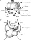
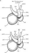
Usually, histological demonstration of the human sinus venosus did not include the IVC (e.g., Blom et al. [6]). Moreover, when the researchers wished to show the IVC terminal, photographic demonstrations of the venous pole of the heart were consistently provided by frontal or sagittal sections [1, 2, 7, 8]. Therefore, it seemed to be still unclear which structure is adjacent to the IVC terminal: the right atrium, left atrium or both. In particularly, the topographical relation of the left and right sinus venosus horns (RSVHs) with the IVC might not be demonstrated in contrast to rather simple diagrams in textbooks of human embryology (Fig. 1): do the three venous elements join together to open the right atrium? In the present study, according to Anderson et al. [1] and Sizarov et al. [2], we called the RSVH to a terminal course of the IVC after joining the left sinus venosus horn. Likewise, in the larger specimens without the left sinus horn, we will use the term for the upper continuation of the IVC in the upper side of the coronary sinus opening. Consequently, in the present study, we attempted to examine serial horizontal sections including not only the heart but the liver to reveal topographical anatomy of the embryonic venous pole of the heart.

Materials and Methods

The study was performed in accordance with the provisions of the Declaration of Helsinki 1995 (as revised in Edinburgh 2000). Horizontal serial paraffin sections stained with hematoxylin and eosin had been obtained from 15 human embryos of crown-rump length (CRL) 9-31 mm, corresponding to gestational ages of approximately 6-7 weeks or Carnegie stage 14-16 [2, 8]. All specimens were part of the large collection kept at the Embryology Institute, Universidad Complutense, Madrid, and were products of miscarriages and ectopic pregnancies managed at the Department of Obstetrics at the university. The study protocol was approved by our university ethics committee (No. B08/374). Our previous studies [9, 10] used heads, necks and abdomens of the same embryos without any anomalies.

Results

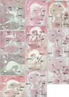
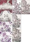
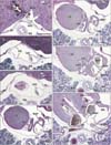
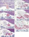
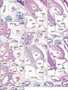
We identified the left sinus venosus horn in 5 of the 15 specimens: the specimen size or CRL was 9 mm, 10 mm, 11 mm, 14 mm, and 15 mm (Figs. 2, 3). Conversely, in specimens larger than 16 mm CRL, we observed the coronary sinus draining into the right atrium (Figs. 4, 5, 6). The IVC terminal and its upper continuation (i.e., the RSVH) consistently provided a thick, continuous tube-like structure. Much or less extent, the IVC was not attached to any part of the heart and liver (Figs. 2C, 3B, 4B, 4C, 5C, 6A). Especially in 3 of the 15 embryos, a part of the course was absolutely free in the pericardial cavity (Fig. 5C). In 9 of the 15 embryos (Figs. 3B, 3C, 4F, 5D, 5E, 6C, 6D), the IVC was connected with the developing right pulmonary vein by a mesentery-like fibrous tissue. The wall of the IVC was thin near the liver, but, during its upward course, muscular elements of the sinus venosum or right atrium appeared to join the IVC (Figs. 3C, 4D). The caudal end of the sinus was separated from the IVC and free in the pericardial space in 2 embryos (Fig. 3B).
The right atrium was consistently attached to the ventral aspect of the IVC and RSVH along a relatively long course (more than 1 mm in Fig. 6). The right auricle or appendage was seen ballooning from the cranial part of the atrial wall (Figs. 2J, 2K, 3E, 3F, 4G, 6E, 6F). Notably, depending on regression of the left sinus venosus horn, instead, the left atrium appeared to the left side and adjacent to the IVC (Figs. 5D, 6C). The left atrial cavity was separated by the left sinus valve from the RSVH (Figs. 5F, 6E). Thus, the left sinus valve appeared to be the interatrial septum, but we discriminated the valve from the septum according to observations of the serial sections. The septum primum was seen in the ventral and left sides of the left sinus valve (Figs. 2K, 3G, 3H), while a candidate of the septum secundum was seen near and above the ventral attachment of the left sinus valve (Fig. 6G). Before development of the septum primum, the left sinus valve was a definite septal structure between the RSVH and left atrium. The site at which the bilateral pulmonary veins joined was consistently separated from the RSVH by the left sinus valve. When the pulmonary veins reached the heart, the left sinus valve became evident.
The IVC and RSVH were mostly or partly surrounded by the left and right venous valves, but the opening of the RSVH to the atrium was located, in 14 of the 15 specimens, on the far caudal side of the opening of the right common cardinal vein, with the distance between the caval vein terminals ranging from 0.3-0.7 mm. Thus, almost always, both the IVC and the future superior vena cave were not seen in the same section. One exception is shown in Fig. 2, in which both caval veins were joined in a panel (Fig. 2H). Because the sinus valve was better developed on the right than on the left side, the RSVH in 8 specimens opened to the left atrium or pulmonary vein before (in the lower side of) opening to the right atrium (Figs. 3G, 6G). In one single specimen, the RSVH opened bilaterally in a single section (Fig. 5G). Because the IVC and RSVH were absolutely continuous, we did not find a reason why we had to discriminate the RSVH from the IVC in the upper side of the coronary sinus opening. Therefore, the IVC (not the RSVH) was likely to open the left atrium in the lower side of the right-sided opening because of the left sinus valve much smaller than the right one.
Consequently, rather than the future superior cava, the IVC and RSVH were a major provider of a right-sided atrial lumen at 6-7 weeks. The observations were summarized in Fig. 7.

Discussion

We found that the right atrium extended far caudally along the IVC at stages in which a lumen of the IVC and RSVH occupied in most or major parts of the right atrial structure. In contrast to a fold-like scheme provided by Blom et al. [6], the right sinus valve absolutely separated most of the long IVC-RSVH course from the right atrium. Thus, we suspected almost complete absorption of the right sinus valve to provide the smooth-walled atrium. Instead, another possibility seemed to be likely that, in the later stage, the growth or elongation of the IVC is much smaller than other parts of the right atrium. The IVC was often fixed to the right pulmonary vein by a mesentery-like fibrous tissue. The IVC developing in the pericardial cavity earlier than the pulmonary vein development may provide a traction to make the so-called pulmonary pit for the joining of the later veins. Actually, a developing vessel is likely to make a thick fascial sheath in human fetuses [11]. This fibrous fixation may explain the pathogenesis of the scimitar anomaly [12].
The usual diagram including the large primitive atrium connected with the sinus venosus (Fig. 1) (e.g., Sadler [13], Moore and Persaud [14]) seemed to be unlikely because the atrium extended inferiorly along the IVC. The IVC and RSVH were as thick as the early atrial cavities (Fig. 7). Moreover, the left atrium was likely to be adjacent to the IVC in the present specimens of CRL 9-31 mm. The simplest explanation was found in a stage-dependent change, with the left atrium developing caudally to separate the sinus venosus or coronary sinus from the IVC and RSVH during different stages. However, the typical figure of the left and right sinus valves surrounding the IVC in the right atrium, such as shown in previous studies [1, 7], seemed to be a rather rare morphology or a product chosen in frontal or sagittal sections. Although the "stages" are believed to determine any significant difference in topographical relation in the heart, depending on individual variations, the left sinus valve was likely to separate the IVC from the left atrium. Likewise, we found that, along the long course, the IVC was attached to the dorsal aspect of the right atrium. In adult hearts, the IVC wall attaches to the right atrium at a wide area, while the former attaches to the left atrium at the right posterior margin. We considered that these attachment sites were derived from the right and left sinus valves, respectively. This hypothesis did not contradict with the incorporation theory of the sinus venosus into the right atrial wall [7].
In conclusion, instead of the so-called primitive atrium (Fig. 1), the IVC and RSVH were a major provider of a right-sided atrial lumen at 6-7 weeks (Fig. 7). Variations in topographical anatomy around the IVC, especially of the sinus valves, might not always depend on the stages but partly in individual differences.

Figures and Tables

Fig. 1
Schematic representations of the early fetal atrium and venous routes. Dorsal views. In panels (A) and (B), a structure large as the ventricles (LV, left ventricle; RV, right ventricle), termed the primitive atrium, is evident in the ventral side of the sinus venosus horns (left sinus venosus horn [LVSH], right sinus venosus horn [RVSH]) and inferior vena cava (IVC). LCCV, left common cardinal vein; RCCV, right common cardinal vein.
acb-47-236-g001
Fig. 2
Inferior vena cava terminal in an embryo of crown-rump length 10 mm. Panels A and K show the most caudal and cranial sides of the figure, respectively. The intervals between panels were 0.05 mm (A, B), 0.16 mm (B, C), 0.1 mm (C, D), 0.06 mm (D, E), 0.02 mm (E, F), 0.01 mm (F, G), 0.14 mm (G, H), 0.06 mm (H, I), 0.16 mm (I, J), and 0.1 mm ( J, K). A part of the sinus venosus (SV) was observed on the left side of the inferior vena cava (IVC) (D, E), communicating with the left horn (LSVH) (arrows in panels F and G). The left sinus horn extended cranially (H, I), continuing to the left common cardinal vein (LCCV) ( J). The right sinus venosus horn (RSVH) opened to the right atrium (RAT) (K) 0.35 mm to the cranial side of the opening of the right common cardinal vein (RCCV) or superior vena cava (H). ES, esophagus; LHV, left hepatic vein; LL, left lung ; LSV, left sinus valve; LSVH, left sinus venosus horn; LV, left ventricle; MHV, middle hepatic vein; PC, pericardium; RA, right auricle; RL, right lung; RSV, right sinus valve; RV, right ventricle; SP, septum primum; TR, trachea. All panels were prepared at the same magnification. Scale bar=1 mm.
acb-47-236-g002
Fig. 3
Inferior vena cava terminal in an embryo of crown-rump length 16.5 mm. Panels (A) and (G) show the most caudal and cranial sides of the figure, respectively. The intervals between panels were 0.5 mm (A, B), 0.05 mm (B, C), 0.15 mm (C, D), 0.05 mm (D, E), 0.1 mm (E, F), and 0.8 mm (F, G). A part of the sinus venosus (SV) was present on the left side of the inferior vena cava (IVC) (D, E) and communicated with the coronary sinus (CS) (F). The right atrium (RAT) was attached to the ventral aspect of the IVC (D, E). Communication between the right sinus venosus horn (RSVH) and the right atrium (G) was located in 0.6 mm caudal side of the right common cardinal venous opening (not shown). The arrow in panel (F) indicates a fibrous connection between the right pulmonar y ve in (RPV ) and the IVC. AO, aorta; DI, diaphragm; ES, esophagus; LAT, left atrium; LHV, left hepatic vein; LL, left lung; LPV, left pulmonary vein; LSV, left sinus valve; LV, left ventricle; MHV, middle hepatic vein; PC, pericardium; RA, right auricle; RAT, right atrium; RL, right lung; RSV, right sinus valve; RV, right ventricle; SP, septum primum; SSE, septum secundum; SV, sinus venosum. All panels were prepared at the same magnification. Scale bar=1 mm.
acb-47-236-g003
Fig. 4
Inferior vena cava terminal in an embryo of crown-rump length 16.5 mm. Panels (A) and (G) show the most caudal and cranial sides of the figure, respectively. The intervals between panels were 0.5 mm (A, B), 0.05 mm (B, C), 0.15 mm (C, D), 0.05 mm (D, E), 0.1 mm (E, F), and 0.8 mm (F, G). A part of the sinus venosus (SV) was present on the left side of the inferior vena cava (IVC) (D, E) and communicated with the coronary sinus (CS) (F). The right atrium (RAT) was attached to the ventral aspect of the IVC (D, E). Communication between the right sinus venosus horn (RSVH) and the right atrium (G) was located in 0.6 mm caudal side of the right common cardinal venous opening (not shown). The arrow in panel (F) indicates a fibrous connection between the right pulmonar y ve in (RPV ) and the IVC. AO, aorta; DI, diaphragm; ES, esophagus; LAT, left atrium; LHV, left hepatic vein; LL, left lung; LPV, left pulmonary vein; LSV, left sinus valve; LV, left ventricle; MHV, middle hepatic vein; PC, pericardium; RA, right auricle; RAT, right atrium; RL, right lung; RSV, right sinus valve; RV, right ventricle; SP, septum primum; SSE, septum secundum; SV, sinus venosum. All panels were prepared at the same magnification. Scale bar=1 mm.
acb-47-236-g004
Fig. 5
Inferior vena cava terminal in an embryo of crown-rump length 21 mm. Panels (A) and (G) show the most caudal and cranial sides of the figure, respectively. Intervals between panels were 0.2 mm (A, B), 0.3 mm (B, C), 0.2 mm (C, D), 0.1 mm (D, E), 0.2 mm (E, F), and 0.4 mm (F, G). The left atrium (LAT) was present on the left side of the inferior vena cava (IVC) (C, D). No left sinus horn was visible. The right atrium (RAT) was attached to the ventral aspect of the right sinus venosus horn (RSVH) (E, F), which opened to the RAT and LAT (G) 0.7 mm to the caudal side of the opening of the right common cardinal vein (not shown). The arrows in panels (D) and (E) indicate a fibrous connection between the right pulmonary vein and the IVC and RSVH. AO, aorta; CS, coronary sinus; DI, diaphragm; ES, esophagus; LA, left auricle; LHV, left hepatic vein; LL, left lung; LSV, left sinus valve; LV, left ventricle; MHV, middle hepatic vein; PC, pericardium; PV, pulmonary veins; RL, right lung; RSV, right sinus valve; RV, right ventricle; SP, septum primum. All panels were prepared at the same magnification. Scale bar=1 mm.
acb-47-236-g005
Fig. 6
Inferior vena cava terminal in an embryo of crown-rump length 22 mm. Panels (A) and (H) show the most caudal and cranial sides of the figure, respectively. The intervals between panels were 0.1 mm (A, B), 0.05 mm (B, C), 0.15 mm (C, D), 0.05 mm (D, E), 0.1 mm (E, F), and 0.8 mm (F, G), respectively. A part of the sinus venosus was present on the left side of the inferior vena cava (IVC) (D, E) and communicated with the coronary sinus (CS) (F). The right atrium (RAT) was attached to the ventral aspect of the IVC (D, E) and communicated with the right sinus venosus horn (RSVH) (H) 0.6 mm to the caudal side of the opening of the right common cardinal vein (not shown). The arrow in panel (F) indicates a fibrous connection between the right pulmonary vein (RPV) and the IVC. CS, coronary sinus; ES, esophagus; GCV, great cardiac vein; LA, left auricle; LAT, left atrium; LL, left lung ; LPV, left pulmonary vein; LSV, left sinus valve; LSVH, left sinus venosus horn; LV, left ventricle; PV, pulmonary veins; RA, right auricle; RL, right lung; RSV, right sinus valve; RV, right ventricle; SSE, septum secundum. All panels were prepared at the same magnification. Scale bar=1 mm.
acb-47-236-g006
Fig. 7
Topographical anatomy of the early inferior vena cava and its surroundings. Dorsal views of embryos with 9-31 mm crown-rump length. Panel (A) displays the initial morphology in which the left atrium has not yet developed, while panel (B) exhibits the developing phase of the left-sided structures. Instead of the primitive atrium (a common chamber), the right atrium (RAT) was as large as the left sinus venosus horn and extend caudally along the inferior vena cava (IVC). Opening of the sinus venosus to the atrium (Fig. 1) corresponds to the upper end of the IVC (or the right sinus venosus horn) that is sandwiched by the left and right sinus valves (LSV, RSV). The developing pulmonary vein (PV) is connected with the dorsal aspect of the IVC by a fibrous structure (star). LAT, left atrium; LCCV, left common cardinal vein; LSVH, left sinus venosus horn; LV, left ventricle; RCCV, right common cardinal vein; RSVH, right sinus venosus horn; RV, right ventricle.
acb-47-236-g007

Acknowledgements

This study was supported by a grant (0620220-1) from the National R & D Program for Cancer Control, Ministry of Health & Welfare, Republic of Korea.

References

1. Anderson RH, Brown NA, Moorman AF. Development and structures of the venous pole of the heart. Dev Dyn. 2006; 235:2–9.
2. Sizarov A, Anderson RH, Christoffels VM, Moorman AF. Three-dimensional and molecular analysis of the venous pole of the developing human heart. Circulation. 2010; 122:798–807.
3. Dickson AD. The development of the ductus venosus in man and the goat. J Anat. 1957; 91:358–368.
4. Mavrides E, Moscoso G, Carvalho JS, Campbell S, Thilaganathan B. The anatomy of the umbilical, portal and hepatic venous systems in the human fetus at 14-19 weeks of gestation. Ultrasound Obstet Gynecol. 2001; 18:598–604.
5. Collardeau-Frachon S, Scoazec JY. Vascular development and differentiation during human liver organogenesis. Anat Rec (Hoboken). 2008; 291:614–627.
6. Blom NA, Gittenberger-de Groot AC, Jongeneel TH, DeRuiter MC, Poelmann RE, Ottenkamp J. Normal development of the pulmonary veins in human embryos and formulation of a morphogenetic concept for sinus venosus defects. Am J Cardiol. 2001; 87:305–309.
7. Steding G, Xu JW, Seidl W, Männer J, Xia H. Developmental aspects of the sinus valves and the sinus venosus septum of the right atrium in human embryos. Anat Embryol (Berl). 1990; 181:469–475.
8. Webb S, Kanani M, Anderson RH, Richardson MK, Brown NA. Development of the human pulmonary vein and its incorporation in the morphologically left atrium. Cardiol Young. 2001; 11:632–642.
9. Hayashi S, Fukuzawa Y, Rodríguez-Vázquez JF, Cho BH, Verdugo-López S, Murakami G, Nakano T. Pleuroperitoneal canal closure and the fetal adrenal gland. Anat Rec (Hoboken). 2011; 294:633–644.
10. Rodríguez-Vázquez JF, Kim JH, Verdugo-López S, Murakami G, Cho KH, Asakawa S, Abe S. Human fetal hyoid body origin revisited. J Anat. 2011; 219:143–149.
11. Miyake N, Hayashi S, Kawase T, Cho BH, Murakami G, Fujimiya M, Kitano H. Fetal anatomy of the human carotid sheath and structures in and around it. Anat Rec (Hoboken). 2010; 293:438–445.
12. Bartram U, Van Praagh S, Keane JF, Lang P, van der Velde ME, Van Praagh R. Mitral and aortic atresia associated with hypoplastic right lung, crossover segment of right lower lobe, and anomalous scimitar-like right pulmonary venous connection with inferior vena cava: clinical, angiocardiographic, and autopsy findings in a rare case. Pediatr Dev Pathol. 1998; 1:413–419.
13. Sadler TW. Langman's medical embryology. 7th ed. Baltimore: Williams & Wilkins;1995.
14. Moore KL, Persaud TV. The developing human: clinically oriented embryology. 6th ed. Philadelphia: W. B. Saunders;1998.
TOOLS
Similar articles