

 PDF
PDF ePub
ePub Citation
 Print
Print


Introduction
Normally the right kidney is supplied by a single renal artery (RA), which is a lateral branch of the abdominal aorta. Near the hilum of the kidney, the RA divides into anterior and posterior divisions. These divisions of RA enter the kidney through the posterior part of the hilum, behind the renal pelvis. The renal vein emerges out through the anterior part of the hilum, in front of the renal pelvis [1]. The RA can vary in its number, point of origin, course or branching pattern. When we have more than one RA, the arteries other than the main RA are called accessory renal arteries. Branches of RA can enter the kidney through the hilum or through the surfaces or poles of the kidney. The branches that enter the kidney through the surfaces are called aberrant renal arteries. We report a rare branching pattern of the right RA. It gave eight prehilar branches which entered the kidney in and around the hilum. We discuss the functional and clinical importance of this variation.
Case Report
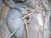
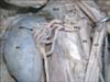
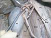
During routine dissections, a variation in the branching pattern of the right RA was noted in an adult male cadaver aged approximately 70 years. The right RA first divided into upper and lower divisions behind the inferior vena cava, 6 cm away from the hilum of the right kidney. These two divisions had a criss-cross arrangement (Figs. 1, 2). The upper division gave 4 renal branches, 3 cm away from the hilum of the kidney. It also gave origin to the right inferior suprarenal artery. The lower division gave two branches, 5 cm away from the kidney. These two branches further bifurcated and gave 2 branches each 3-4 cm away from the hilum. Thus, there were 8 prehilar branches of RA (Figs. 1, 2). The branches of the upper division occupied the anterior part of the renal hilum and the branches of the lower division occupied the posterior part of the renal hilum (Fig. 2). The upper two among the four branches of the lower division entered the kidney through its anterior surface, just above the hilum, thus being aberrant arteries. The renal pelvis occupied the centre of the hilum. The right renal vein emerged from the uppermost part of the hilum and had a normal course and termination. The multiple prehilar branches led to a congested atmosphere at the hilum of the kidney (Fig. 3).
Discussion
Though there are many reports on the accessory and aberrant renal arteries, reports on multiple branches of the single RA are scanty. Occurrence of accessory renal arteries is mainly due to the failure of degeneration of caudal mesonephric arteries during the developmental stages of the kidney [2]. There is no clear explanation available in the literature about the exact cause of early division of the RA. According to Bayramoglu et al. [3] the variations in the number of renal arterial divisions in the hilar region are generally associated with renal malformations in the embryo. Ozkan et al. [4], in their angiographic study on 855 patients, noted early division of RA only in 8% of cases. Nayak [5] has reported a case where there were three right renal arteries. The superior RA gave three branches; middle and inferior gave two branches each. Among the seven branches, two of them pierced the anterior surface of the kidney. The current case had only one RA which divided into two divisions and each of the divisions gave 4 prehilar branches. In the case reported by Nayak [5] the right inferior suprarenal artery arose from the middle RA, whereas in the current case it was a branch of the upper division of the RA. In a case reported by Kumar et al. [6] the right RA divided into upper and lower divisions. The upper division pierced the upper pole of the kidney and the inferior division gave 6 branches; four of which entered the kidney through the hilum, in front of the renal pelvis and the remaining two passed behind the renal pelvis. The current case differs from above case in having equal number of branches from upper and lower divisions. Apart from this, there was a crisscross arrangement between the two divisions of the RA.
The early division of the right RA, the crisscross arrangement of the two divisions and eight branches from two divisions might lead to hindrance in the flow of blood to the kidney due to the vascular congestion. It might also lead to compression of the renal vein or the renal pelvis. Rouviere et al. [7] observed 29-65% incidence with anomalous course of renal vessels crossing the renal pelvis associated with ureteropelvic obstruction. Anomalous position of the vessels in the renal hilum is believed to be because of the rotational defects of the kidney [8]. Anatomical knowledge of the number and arrangement of the structures in the renal hilar region is important for a number of radiological and surgical procedures. Surgical intervention that requires renal hilar dissection needs separate clamping of the vessels and renal pelvis which is preferred over en bloc mass stapling of renal hilum. Presence of too many structures at the hilum might make hilar dissection difficult and might convert the conversion of laparoscopic operation to an open procedure [9]. Knowledge of the variations reported here is important while performing endovascular procedures like therapeutic embolisation and angioplasties. It is also important in nephron-preserving surgery, kidney transplantation, and the management of renal vascular hypertension. Since these variations can be demonstrated preoperatively by selective angiography, the awareness of this variation is of utmost importance to the radiologists.
 XML Download