

 PDF
PDF ePub
ePub Citation
 Print
Print


Introduction
Ganglion cysts are mucin filled cystic lesions associated with the synovium of either a joint or a tendon sheath. It commonly occurs on the dorsum of the hand, wrist and foot. The cyst may have a single cavity or multilobulated. Cysts in the popliteal fossa which communicate with the knee joint by means of a stalk, are called Baker's cysts. Non-communicating cysts are ganglion cysts, juxta articular myxomas [1]. The following is the internationally second ever reported case of an intratendinous ganglion cyst arising from the semimembranosus tendon, with the first being reported in 2009 [2].
Case Report
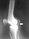
During the dissection of popliteal fossa in a cadaver of a 64-year-old male, a grey white cystic swelling of 2.5 cm×2 cm×0.5 cm was observed attached to the tendon of the right semimembranosus, 3.5 cm above the insertion of the muscle (Fig. 1). A transparent gelatinous fluid was aspirated from the mass, indicating a cyst. Thickness of the smooth lining cyst wall was 0.2 cm. The stalk of the cyst was seen. To ascertain its communication with the knee joint, radio-opaque dye was injected. Contrast X-ray revealed it as non-communicating cyst (Fig. 2). Histopathology of the cyst revealed: cyst wall showing fibro collagenous tissue, lined by cuboidal epithelium. Adjacent soft tissue shows fibro collagenous tissue and adipose tissue with foci of myxoid and hyaline degeneration (Fig. 3).
Discussion
Ganglion cyst is a common tumor of hand and wrist. Sixty to seventy percent arise on the dorsal aspect of the wrist and remaining 20% on the volar aspect [3]. It commonly occurs in the 2nd to 4th decade of life. Exact cause of the Ganglion cyst is not clear, and is usually multifactorial. Both local stress and congenital anomalous origin have been ascribed. Various theories of origin are mucinous degeneration of fibrous tissue, or a herniation of synovium secreting hyperactively as a nonspecific synovitis, or a retention cyst. It can arise from stress due to stretching of capsular and ligamentous structures, which stimulates the production of mucin from modified synovial, mesenchymal and fibroblast cells that produce hyaluronic acid. Mucin dissects through the capsule and ligamentous tissues forming main cyst. The fluid may enter the cyst from capsular ligamentous interface through a one-way valve type of mechanism and then decrease as the water component is reabsorbed producing a fluctuating cyst. Mucinous fluid is rich in hyaluronic acid, glycosamine, albumin and globulin [3].
The size of the cyst may fluctuate with activity of the affected part. It may resolve by regression or rupture. The cyst may present with dull ache or paresthesia with compression of adjacent nerves. Large cysts can fluctuate and transilluminate. Differential diagnosis for mass in popliteal fossa is popliteal vein varices, popliteal artery aneurysm, synovial sarcoma, haematoma or abscess [3].
Ultrasound and magnetic resonance imaging can differentiate a ganglion cyst from other soft-tissue tumors and tumor-like lesions, and provide excellent information on the location of an intratendinous lesion [2]. Treatment is empirical, by aspiration of the cyst with or without injection of steroid [4]. Excision of the cyst is sometimes followed by recurrence. Recurrence rates range from 4% to 40% [5]. Any cystic mass found in popliteal fossa during operative exploration, can thus be communicating with joint capsule or the semimembranosus tendon.


 XML Download