Journal List > Anat Cell Biol > v.47(3) > 1071776

Lee, Kim, Lee, Park, Jeong, Lee, Han, Kim, and Han: Sex determination using upper limb bones in Korean populations

Abstract

The purpose of this research is to establish metric standards for the determination of sex from the upper limb bones of Korean. We took a set of eleven measurements on each of 175 right sides of adult skeletons chosen at Korean sample. Classification accuracy dropped only one or two individuals when only vertical head diameter of humerus is used. Variables in relation with maximal length were less accurate than head diameter of humerus. Two variables were selected by the stepwise procedure: maximal length of humerus, vertical head diameter of humerus. The combined accuracy was 87%. This study of modern Korean skeletons underscores the need for population-specific techniques, not only for medicolegal investigations, but also for the study of population affinities and factors affecting bone configurations.

Introduction

Sex determination of unidentified skeletal remains from crime scenes or excavation sites is an important component for identification in forensic anthropology. Of the human skeleton, the humerus often remains in good condition and is especially favorable for metric sex determination [1]. The length of the humerus, among the long bones of the human body, is a good predictor, but the vertical head diameter of this bone is also an accurate predictor of gender [2, 3]. For this reason, numerous studies and metric methods have emerged from upper limb bones (ULB) with humerus analyses based in Varanasi [4], prehistoric central California [1], the Terry collection [5], China, Japan, Thailand [6], South Africa [7, 8], Spain [9], Germany [10], Guatemala [11], the Dart collection [11, 12], the island of Crete [2, 13, 14], Chile [3], Turkey [15], Greece [16], America [17], and the Eastern Adriatic coast [18]. Non-metric studies involving the ULB have also been performed in Canada [19] and Japan [20].
Interestingly, the size of the ULB varies distinctly by regional population. For example, humerus lengths are different between the European and American populations. Asians, too, have been shown to exhibit significant regional differences [6]. Within various Asian populations, the discriminant value of humerus size has been determined for the Chinese, Japanese, and Thai populations [6]. However, humerus, radius, and ulna values have yet to be determined for the Korean population. Sex determination reports have been published for the pelvic bone [21], skull [22, 23], corpus callosum [24, 25], hyoid bone [26], thyroid cartilage [27], and mandible [28, 29] in Korean sample populations.
Involving only fragmentary or incomplete remains, sex can be determined by quantitative metric analysis, although with somewhat lower accuracy. Additionally, the ULB unearthed in the forensic field are often fragmented. Thus, the discriminating values from such ULB have been derived from not only complete bone, but also fragmented humeri in Korea. Therefore, the purpose of this research is to establish accurate metric standards for sex determination based on humerus, radius and ulna measurements in the Korean population, as well as to compare the size and sexual dimorphism to studies from other regions.

Materials and Methods

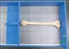
The ULB lengths and partial bone lengths were measured for 175 right side of adult Korean cadavers (100 men, 75 women) between 42 to 95 years of age (mean, 72.2 years; standard deviation, 13.7 years). The ULB were removed from fresh cadavers. Articulate cartilage was removed using a surgical knife. No bones with obvious pathologies or healed fractures were included, and all measurements were taken using an osteometric table (EGO, Seoul, Korea), digital calipers (Mitutoyo, Tokyo, Japan) and measuring tape (Komelon, Busan, Korea) (Fig. 1).
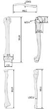
Eleven variables were taken: maximum length of clavicle includes distance between acromial end and sterna end, circumference at middle of shaft of clavicle (CMS), maximum length of humerus includes distance between trochlea and the proximal extremity of humeral head (MLH), maximum diameter of humeral head (MDH), epicondylar breadth of humerus (EB), condylar breadth of humerus, transverse diameter of humeral head (TDH), vertical diameter of humeral head (VDH), maximum length of radius includes distance between styloid process and the proximal extremity of radial head, maximum length of ulna includes distance between styloid process and the proximal extremity of olecranon, least cricumference of ulna shaft (LCSU) (Table 1, Fig. 2).
Statistical analysis was performed using SPSS ver. 17.0 (SPSS Inc., Chicago, IL, USA) for Windows. Means and standard deviations of the results of individual factors were calculated and statistically analyzed to identify any significant differences.

Results

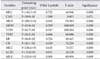
Table 2 represents the mean length and standard deviation of Korean ULB for each of the given variables. All of the values were larger for males than for females, and these differences were statistically significantly. The absolute values for all variables ranged from 3.0 mm to 24.8 mm between males and females.
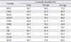
Table 3 presents the discriminant function coefficient of the ULB dimensions for the Korean sample. The functions are displayed based on a single variable. Females are indicated when the discriminant score is lesser than the demarcation point, and males are indicated when the discriminant score is higher. For example, a VDH size of 45 mm would be identified as a male because the diameter is greater than the 42.7 mm function coefficient.
In Table 4, the accuracy of sex determination for the ULB size is represented. In general, the value for humerus breadth was a better predictor than the value of humerus length. The best predictor for sex determination is VDH (87.0%) and the worst predictive value is CMS (60.3%). Specifically, the variables for humeral head including (MDH, VDH, and TDH) were predictive for sex.
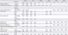
Table 5 depicts a stepwise discriminant analysis of the variables for MLH and VDH. The cutoff point was set to zero. If the product of the predictor variable and its coefficient are added to the constant and yield a value of >0, the individual is classified as male, whereas a sum of <0 indicates a female.

Discussion

Forensic experts are often faced with a single specimen from which he or she must draw conclusions about the specimen's origin. In some cases, it may even be necessary to establish whether remains belong to a specific person when the identity is suspected based on circumstantial evidence. Multiple studies have shown that the ULB is a useful bone for metric sex determination. Such studies also demonstrate the differences in ULB dimensions between populations, potentially resulting from environmental factors such as nutrition, physical development, and genetic factors [11]. Our study is important because it is the first to analyze sex-specific differences in a Korean population sample.
Previous studies indicate that there are population differences between our Korean sample and other populations. The mean lengths of all variables were similar among the different Asian populations. However, Asian ULB sizes are generally smaller than most other populations, with the exception of Guatemala (Table 6). The MLH study from Steyn and Iscan [7] used a South African white sample population whose dimensions (335.0 mm in males and 309.0 mm in females) are larger than that of Koreans by approximately 32.0 mm in both sexes. The t-test showed that all measurements used in the present study were significantly higher in men than in women (P<0.05) (Table 6).
A comparison of the results of the present study with those from other studies' skeletal long bones demonstrates that the same variables are especially valuable. In the LCSU, Spain [9] attained 91.1% accuracy, while our study achieved 60.3% accuracy. Thus, LCSU is a better predictor of sex in the Spanish study sample than the Korean sample (Table 7).
In the other studies, the classification rate is variable: 79.5-89.5% in Prehistoric Central California; 77.9-93.3% in Chinese, Japanese, and Thai populations; 84.0-91.0% in South African white and black populations; 80.6-90.4% in Germans; 83.0-95.5% in Guatemalans; 85.1-89.9% in Cretans; 87.0% in Chileans; and 74.7-87.0% in Koreans. The general effective single variables, as determined by direct discriminant analysis of all population, were VDH and EB except EB of Korea.Additionally, these 2 variables were significantly different between populations (Table 8).
The demarcation points of South African whites were bigger than those determined by other studies. However, the demarcation points of the Eastern Adriatic Coast sample were similar. These differences between populations could be the result of environmental factors affecting bone growth, such as nutrition, physical development, or genetic factors (Table 9).
In conclusion, we describe a specific standard for sex determination in the Korean population using ULB. This study of modern Korean skeletons underscores the need for population-specific techniques, not only for medicolegal investigations, but also for the study of population-specific attributes and factors affecting bone characteristics.

Figures and Tables

Fig. 1
The measurement method using osteometric table. LAT, lateral; SUP, superior.
acb-47-196-g001
Fig. 2
Variables used for sex determination. CB, condylar breadth of humerus; CMS, circumference at middle of shaft of clavicle; EB, epicondylar breadth of humerus; LCSU, least cricumference of ulna shaft; MDH, maximum diameter of humeral head; MLC, maximum length of clavicle includes distance between acromial end and sterna end; MLH, maximum length of humerus includes distance between trochlea and the proximal extremity of humeral head; MLR, maximum length of radius includes distance between styloid process and the proximal extremity of radial head; MLU, maximum length of ulna includes distance between styloid process and the proximal extremity of olecranon; TDH, transverse diameter of humeral head; VDH, vertical diameter of humeral head.
acb-47-196-g002
Table 1
The measurement of humerus, radius and ulna
acb-47-196-i001
Table 2
Means±SD of measured variables
acb-47-196-i002

Values are presented as mm. MLC, maximum length of clavicle; CMS, circumference at middle of shaft of clavicle; MLH, maximum length of humerus; MDH, maximum diameter of humeral head; VDH, vertical diameter of humeral head; TDH, transverse diameter of humeral head; EB, epicondylar breadth of humerus; CB, condylar breadth of humerus; MLU, maximum length of ulna; LCSU, least cricumference of ulna shaft; MLR, maximum length of radius.

Table 3
Demarking point separating males from females
acb-47-196-i003

MLC, maximum length of clavicle; F, female; M, male; CMS, circumference at middle of shaft of clavicle; MLH, maximum length of humerus; MDH, maximum diameter of humeral head; VDH, vertical diameter of humeral head; TDH, transverse diameter of humeral head; EB, epicondylar breadth of humerus; CB, condylar breadth of humerus; MLU, maximum length of ulna; LCSU, least cricumference of ulna shaft; MLR, maximum length of radius.

Table 4
Percentage of correctly classified cases
acb-47-196-i004

MLC, maximum length of clavicle; CMS, circumference at middle of shaft of clavicle; MLH, maximum length of humerus; MDH, maximum diameter of humeral head; VDH, vertical diameter of humeral head; TDH, transverse diameter of humeral head; EB, epicondylar breadth of humerus; CB, condylar breadth of humerus; MLU, maximum length of ulna; LCSU, least cricumference of ulna shaft; MLR, maximum length of radius.

Table 5
Stepwise discriminant function analysis
acb-47-196-i005

MLH, maximum length of humerus; VDH, vertical diameter of humeral head.

Table 6
Comparison mean length of each variables with other studies
acb-47-196-i006

MLH, maximum length of humerus; EB, epicondylar breadth of humerus; TDH, transverse diameter of humeral head; VDH, vertical diameter of humeral head; MLR, maximum length of radius; MLU, maximum length of ulna; M, male; F, female.

Table 7
Comparison Safont et al. [9] with this study about percentage of correctly
acb-47-196-i007

LCSU, least cricumference of ulna shaft.

Table 8
Comparison of percentage of sex determination with single measurements
acb-47-196-i008

Values are presented as percentage. TDH, transverse diameter of humeral head; VDH, vertical diameter of humeral head; EB, epicondylar breadth of humerus; MLH, maximum length of humerus; MLU, maximum length of ulna; MLR, maximum length of radius.

Table 9
Comparsion of demarking point with other studies
acb-47-196-i009

MLH, maximum length of humerus; VDH, vertical diameter of humeral head; EB, epicondylar breadth of humerus; F, female; M, male.

Acknowledgements

We thank Hong Byung Ook and Kim Sang Hyun, Department of Anatomy, Catholic Institute for Applied Anatomy, The Catholic University of Korea College of Medicine, for help in gathering the data.
This research was supported by basic science research program through the national research foundation of Korea (NRF) funded by the Ministry of Education, Science and Technology (No. 2014R1A1A1006195).

References

1. Dittrick J, Suchey JM. Sex determination of prehistoric central California skeletal remains using discriminant analysis of the femur and humerus. Am J Phys Anthropol. 1986; 70:3–9.
2. Kranioti EF, Bastir M, Sánchez-Meseguer A, Rosas A. A geometric-morphometric study of the Cretan humerus for sex identification. Forensic Sci Int. 2009; 189:111.e1–111.e8.
3. Ross AH, Manneschi MJ. New identification criteria for the Chilean population: Estimation of sex and stature. Forensic Sci Int. 2011; 204:206.e1–206.e3.
4. Singh S, Singh SP. Identification of sex from the humerus. Indian J Med Res. 1972; 60:1061–1066.
5. Holman DJ, Bennett KA. Determination of sex from arm bone measurements. Am J Phys Anthropol. 1991; 84:421–426.
6. Işcan MY, Loth SR, King CA, Shihai D, Yoshino M. Sexual dimorphism in the humerus: a comparative analysis of Chinese, Japanese and Thais. Forensic Sci Int. 1998; 98:17–29.
7. Steyn M, Işcan MY. Osteometric variation in the humerus: sexual dimorphism in South Africans. Forensic Sci Int. 1999; 106:77–85.
8. Vance VL, Steyn M, L'Abbé EN. Nonmetric sex determination from the distal and posterior humerus in black and white South Africans. J Forensic Sci. 2011; 56:710–714.
9. Safont S, Malgosa A, Subirà ME. Sex assessment on the basis of long bone circumference. Am J Phys Anthropol. 2000; 113:317–328.
10. Mall G, Hubig M, Büttner A, Kuznik J, Penning R, Graw M. Sex determination and estimation of stature from the long bones of the arm. Forensic Sci Int. 2001; 117:23–30.
11. Ríos Frutos L. Metric determination of sex from the humerus in a Guatemalan forensic sample. Forensic Sci Int. 2005; 147:153–157.
12. Barrier IL, L'Abbé EN. Sex determination from the radius and ulna in a modern South African sample. Forensic Sci Int. 2008; 179:85.e1–85.e7.
13. Kranioti EF, Nathena D, Michalodimitrakis M. Sex estimation of the Cretan humerus: a digital radiometric study. Int J Legal Med. 2011; 125:659–667.
14. Kranioti EF, Michalodimitrakis M. Sexual dimorphism of the humerus in contemporary Cretans: a population-specific study and a review of the literature. J Forensic Sci. 2009; 54:996–1000.
15. Uzün I, Işcan MY, Celbiş O. Forearm bones and sexual variation in Turkish population. Am J Forensic Med Pathol. 2011; 32:355–358.
16. Charisi D, Eliopoulos C, Vanna V, Koilias CG, Manolis SK. Sexual dimorphism of the arm bones in a modern greek population. J Forensic Sci. 2011; 56:10–18.
17. Milner GR, Boldsen JL. Humeral and femoral head diameters in recent white American skeletons. J Forensic Sci. 2012; 57:35–40.
18. Bašić Ž, Anterić I, Vilović K, Petaros A, Bosnar A, Madžar T, Polašek O, Anđelinović Š. Sex determination in skeletal remains from the medieval Eastern Adriatic coast - discriminant function analysis of humeri. Croat Med J. 2013; 54:272–278.
19. Rogers TL. A visual method of determining the sex of skeletal remains using the distal humerus. J Forensic Sci. 1999; 44:57–60.
20. Tanaka H, Lestrel PE, Uetake T, Kato S, Ohtsuki F. Sex differences in proximal humeral outline shape: elliptical Fourier functions. J Forensic Sci. 2000; 45:292–302.
21. Choi BY, Chung IH. Sex discrimination with the metric measurements of the Korean dried pelvic bones by discriminant function analysis. Korean J Phys Anthropol. 1999; 12:151–158.
22. Choi BY, Lee KS, Han SH, Park DK, Lim NH, Koh KS, Kim HJ, Kang HS. Group analysis using the metric measurements of Korean skulls. Korean J Phys Anthropol. 2001; 14:207–215.
23. Park DK, Lee UY, Lee JH, Choi BY, Koh KS, Kim HJ, Park SJ, Han SH. Non-metric traits of Korean skulls. Korean J Phys Anthropol. 2001; 14:117–126.
24. Ji EK, Rhyu IJ, Cheon YH, Lee EK, Kim YM, Shin DY, Hwang SJ. Gender difference of corpus callosum in Korean neonate. Korean J Phys Anthropol. 2001; 14:333–338.
25. Rhyu IJ, Ryu JY, Park KW, Lee DH, Park KY, Park KH, Hwang SJ. Gender difference on corpus callosum in Korean adults. Korean J Phys Anthropol. 2005; 18:169–177.
26. Kim DI, Lee UY, Park DK, Kim YS, Han KH, Kim KH, Han SH. Morphometrics of the hyoid bone for human sex determination from digital photographs. J Forensic Sci. 2006; 51:979–984.
27. Park DK, Kim DI, Lee UY, Han KH, Kim KH, Han SH. Morphometric analysis of the Korean thyroid cartilage for identification of sex: metric study. Korean J Phys Anthropol. 2003; 16:129–136.
28. Hu KS, Koh KS, Park KK, Kang MK, Chung IH, Kim HJ. Non-metric traits of Korean mandibles. Korean J Phys Anthropol. 2000; 13:161–172.
29. Hu KS, Koh KS, Jung HS, Kang MK, Choi BY, Kim HJ. Physical anthropological characteristics and sex determinative analysis by the metric traits of Korean mandibles. Korean J Phys Anthropol. 2000; 13:369–382.
TOOLS
Similar articles