

 PDF
PDF ePub
ePub Citation
 Print
Print


Introduction
Living donor liver transplantation (LDLT) has been adopted as the preferred method of treatment for adults with end stage liver disease due to shortage of the available cadaveric donors [1]. In LDLT, injury to the hepatic arterial system could lead to severe complications like hepatic artery thrombosis (HAT), which may result in graft loss in the recipient or significant reduction in the blood supply to the remaining liver in the donor or both [2].
Holbert et al. [3] had reported that the segment 4 of the liver is at a greater risk for development of HAT as a post-transplant complication. Michels [4] had defined the artery to the segment 4 of the liver as the middle hepatic artery (MHA). Hence it may be suggested that a detailed knowledge regarding the anatomy of the MHA could have considerable implications in the preservation/reconstruction of the MHA, which could reduce the incidence of HAT as a postoperative complication of LDLT.
The purpose of this paper was to characterize the origin of the MHA in detail and classify the variations observed through the anatomical study of the cadaveric livers, and also to analyze the clinical significance (if any) of the findings in relation to postoperative vascular complications in LDLT.
Materials and Methods
The study was conducted in the Department of Anatomy, Lady Hardinge Medical College and Associated Hospitals, New Delhi, India. The author obtained ethical approval from the Ethics Committee of the above mentioned institution. The livers used in this study were retrieved from human cadavers aged 55-78 years (male, 77; female, 48), obtained from the clinical wards of associated Smt. Sucheta Kriplani Hospital, New Delhi, India. Prior to procurement of the cadavers written informed consent was obtained from the family members/relatives in each case. Only those cadavers, with no history of any liver disease in their medical records, were selected for this study.
Resections were performed very carefully, so as to include the liver, celiac trunk, left gastric artery (LGA), lesser omentum, superior mesenteric artery (SMA) and head of the pancreas, in each of the study specimen. A total of 125 adult livers, without macroscopic abnormalities were examined. According to Couinaud's [5] and Bismuth's [6] liver segmentation, the left medial sub-segment of the liver which lies medial to the falciform ligament, has been defined as the segment 4. Saxena et al. [7] quoting the works of Hjortsjo [8] and Mizumoto and Suzuki [9], had reported that topographically the quadrate lobe constitutes the major part of the left medial sub-segment of the liver. However the quadrate lobe has also been considered as a subdivision of the right anatomical lobe of the liver [10]. Hence it was postulated that the quadrate lobe could possibly be supplied by more than one branch from the hepatic arterial system.
Dissection was started from the inferior aspect of the hepatic hilum and subsequently all the branches of the celiac trunk, SMA and the hepatic arterial system were identified. Meticulous dissection was performed in each specimen and the arterial branches supplying the quadrate lobe of liver were exposed by carefully removing the liver tissue surrounding these branches. The distribution of each arterial branch supplying the quadrate lobe was noted and subsequently the area supplied by these arteries was estimated. For the present study, that branch of the hepatic arterial system which formed the dominant arterial supply of the quadrate lobe of liver was defined as the artery to the hepatic segment 4 or the MHA. Further, an artery arising from the SMA and reaching into the right lobe of the liver was defined as the accessory right hepatic artery (aRHA), and an artery arising from the LGA and reaching into the left lobe of the liver was defined as the accessory left hepatic artery (aLHA).
The MHA was identified in each of the liver specimen, the origin of the artery was traced, and the different variations observed in the origin of the MHA were schematically studied across all the study specimen.
Results
At first the branching pattern of the celiac trunk and the SMA was noted in all the dissected specimens and in 123 cases (98.4%) classical trifurcation of the celiac trunk was observed, whereas in two specimens (1.6%) bifurcation of the trunk was observed (splenogastric trunk) with the common hepatic artery arising from the SMA (hepato-mesenteric trunk) (Fig. 1).
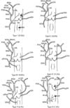
Subsequently the branches of the hepatic arterial system were taken into consideration and in general it was observed that the right hepatic artery (RHA) and the left hepatic artery (LHA) arose as branches of the proper hepatic artery. The LHA divided into two sub-branches; medial and lateral segmental arteries. The medial segmental artery (MSA) supplied the quadrate lobe and anterior region of the left lobe. The lateral segmental artery (LSA) further divided into superior and inferior divisions to supply the left lobe of liver. The inferior division of the LSA also supplied the quadrate lobe. The RHA divided into two sub-branches: anterior and posterior segmental arteries, which further divided into superior and inferior divisions to supply the right lobe of liver. The inferior division of the anterior segmental artery (ASA) also supplied the quadrate lobe of the liver. Hence it was observed that the quadrate lobe in all the livers was supplied by three arteries; MSA and LSA (sub-branches of the LHA) and the ASA (sub-branch of the RHA), with either the MSA or the ASA being the dominant one. Now the MHA was identified in each specimen as the dominant artery which supplied the quadrate lobe of liver and it was observed that the MHA arose as a sub-branch of LHA in fifty-nine specimens (47.2%) and as a sub-branch of RHA in sixty-six specimens (52.8%) (Table 1, Fig. 1).
This was followed by the identification of the accessory hepatic arteries when present in the study specimens. It was noted that the aLHA was present in sixteen cases (12.8%), aRHA in seventeen cases (13.6%) and both the accessory hepatic arteries were present in three cases (2.4%) (Table 1, Fig. 1).
Based on the variations observed in the origin of the MHA, while taking into consideration the presence of accessory hepatic arteries (aLHA/aRHA/both), in the present study the hepatic arterial anatomy was classified into six types (Table 1, Fig. 1). In type I hepatic arterial configuration, the dominant artery to the quadrate lobe was ASA and in type II it was MSA. Type I and type II hepatic arterial configurations were observed in 37.6% and 33.6% cases respectively and they are the normal anatomical pattern observed in most of the adult livers, without the presence of any accessory hepatic arteries. In type III hepatic arterial configuration, the dominant artery to the quadrate lobe was MSA, the aRHA was present and it was observed in 13.6% of livers. In type IV, the dominant artery to the quadrate lobe was ASA, the aLHA was present and it was noted in 11.2% of livers. In type V, the dominant artery to the quadrate lobe was ASA, both the accessory hepatic arteries were present (aRHA and aLHA) and it was observed in 2.4% of the livers. In type VI hepatic arterial configuration, the dominant artery to the quadrate lobe was ASA, common hepatic artery (CHA) was arising from the hepato-mesenteric trunk, aLHA was present and it was noted in 1.6% of the livers (Table 1, Fig. 1).
It was further observed that in seventeen specimens (13.6%) under study, in the presence of aRHA, MHA arose as a sub-branch of LHA (type III in present study) and in nineteen specimens (15.2%), in the presence of aLHA (irrespective of the presence of aRHA), MHA arose as a sub-branch of RHA (type IV to type VI in the present study) (Table 1, Fig. 1).
Discussion
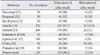
The observations of the present study with regards to the branching pattern of the celiac trunk are different from most of the previous reports (Table 2) [11, 12, 13, 14, 15]. However the findings of the present study are in accordance with the more recent studies like Katsume et al. [16] and Ugurel et al. [17] (Table 2). It is noteworthy that the observations of the present study are different from the findings of Chitra [18] and Prakash et al. [19] which similar to mine were conducted on Indian cadavers (Table 2). Such disparity might be attributed to the difference in the sample size of the cadavers and the difference in the study population groups. In the present study bifurcation of the celiac trunk were observed in two livers (1.6%), where the celiac trunk gave rise to spleno-gastric trunk and the CHA arose from SMA (as the hepato-mesenteric trunk) (Fig. 1). Such variation in the branching of the celiac trunk corresponds to type 5 as per the classification proposed by Adachi [14]. It may be highlighted that my findings regarding variations in the branching of celiac trunk (type VI in this study) (Table 2, Fig. 1) are in accordance with the observations of Hiatt et al. [20] who had reported on the basis of 1,000 cases of liver transplantation, the incidence of hepato-mesenteric trunk (CHA arising from SMA) as 1.5%. Further it may be noted that the incidence of hepato-mesenteric trunk as reported in the present study is close to the findings of Nayak and Vasudeva [21] who had observed that the CHA arose from SMA in 3% cases.
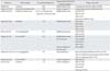
Arterial supply to the hepatic segment 4 was first described by Healey et al. [22] who had reported that the artery was arising in equal proportions from the RHA and the LHA. Michels [4] had defined the artery going to the segment 4 as the MHA that courses in the umbilical fossa and also observed that the artery was arising from RHA and LHA in equal proportions (Table 3). Since then majority of the authors have defined the artery to the segment 4 of the liver as the MHA. Onishi et al. [23] had reported that MHA was arising predominantly from the LHA (61.5%) and in most of the remaining cases from the RHA (27.5%). Futara et al. [24] noted that the MHA was present in 47.3% of cases and was arising in equal proportion from the LHA (20%) and RHA (20%). Kishi et al. [25] observed that MHA was arising from the LHA in 62.5% cases and from the RHA in 37.5% of the cases, which was similar to the findings of Onishi et al. [23]. Wang et al. [26] had defined MHA as the artery which arose from its artery of origin at the hepatic hilum and gave off branch/branches to the hepatic segment 4 at the umbilical fissure. Accordingly they had reported the presence of MHA in 103 cases (71%), and in contrast to previous studies, the MHA arose predominantly from the RHA (58.3%) and in most of the remaining cases from LHA (36.9%) (Table 3). Yoshimura et al. [27] had described the artery to the hepatic segment 4 as the left medial artery (LMA). They had classified the LMA into three types: type I arising from the distal part of the LHA on reaching the umbilical portion of the portal vein (37.2%), type II arising from the proximal part of the LHA before reaching the umbilical portion of the portal vein (35.8%), and type III arising from RHA (27%). Kamel et al. [28] identified the artery supplying the hepatic segment 4 in 39 cases and it arose from the RHA in 62.5% patients. Jin et al. [29] had reported that the artery to segment 4 was arising from the RHA in 53.2% liver specimen, and from the LHA in 32.3% cases (Table 3). To summarize the observations made by different authors in the previous studies, it may inferred that the origin of the MHA showed a high degree of variability and in majority of the cases, the artery was arising from either the LHA or the RHA.
In accordance with the observations made by different authors in the previous studies, it was noted in the present study that the origin of the MHA showed considerable variability and arose from either LHA or the RHA (Table 1, Fig. 1). Moreover the findings of this study were similar to the observations of Kamel et al. [28], Jin et al. [29], and Wang et al. [26] in that the MHA in this study arose predominantly as a branch of RHA (52.8% specimens) and from the LHA (47.2% specimens) in the remaining cases (Table 1, Fig. 1).
The incidence of aLHA arising as a branch of LGA as observed in the present study (Table 4, Fig. 1) is higher than what has been reported previously (Table 4) [20, 30, 31]. However the incidence of aRHA arising as a branch of SMA in the present study is close to recent reports but considerably higher than the findings of Daseler and Anson [30] (Table 4). The occurrence of both aLHA and aRHA as observed in the present study is very much similar to the findings of Hiatt et al. [20] but higher than the observations of Koops et al. [31] (Table 4).
Liver surgery is largely an anatomic exercise and Couinaud [32], a French surgeon and anatomist made significant contributions towards understanding the hepatic arterial configuration. According to Couinaud [33], embryologically there are 3 lobes in the early stage of hepatic formation, each supplied by an embryonic artery of its own. The lateral sector (segment 2) is supplied by the embryonic LHA, the medial and anterior sectors (segments 3, 4, 5, and 8) by the embryonic MHA and the posterior sectors (segments 6 and 7) by the embryonic RHA. The embryonic LHA is derived from LGA, the embryonic MHA from the CHA and embryonic RHA from the SMA (Fig. 2) [33]. In the present study, six types of hepatic arterial configuration were observed and it was noted that the MHA arose as a sub-branch of either RHA or LHA (Table 1, Fig. 1), both of which happens to be the branches of CHA, which happens to be the artery from which MHA arises in the embryonic life [33]. This observation may explain the variations in the origin of MHA, as documented by different authors in previous studies (Table 3) and as observed by the author of the present study (Table 1, Fig. 1).
Saxena et al. [7] reported that the quadrate lobe belongs functionally to the left lobe of the liver, hence during right lobe LDLT, preservation of the LHA and the MHA is critical for adequate blood supply to the remaining left lobe in the donor [34]. Similarly during left lobe LDLT, reconstruction of LHA and MHA, ensures adequate blood supply and thereby viability of the graft [26]. Subsequently it may be suggested that when MHA arises as a sub-branch of LHA in the presence of aRHA (type III in present study) (Table 1, Fig. 1), during either right lobe/left lobe LDLT, the preservation/reconstruction of MHA as per requirement would require no modification in the standard surgical procedure. Thus the risk of intra-operative injury to MHA would be less, thereby reducing the chances of development of post-operative HAT [26]. However when MHA arises as a sub-branch of RHA in the presence of aLHA (type IV to type VI in present study) (Table 1, Fig. 1), during either right lobe/left lobe LDLT, the preservation/reconstruction of the MHA would necessitate a complex surgical technique. During right lobe LDLT, as the right lobe graft would include the RHA, hence it would be very difficult to preserve the MHA in the donor, which is essential for the adequate blood supply and hence survival of the remaining left lobe in the donor. Similarly during left lobe LDLT, the left lobe graft should include the MHA along with the LHA for proper reconstruction of both the arteries and survival of the graft in the recipient. As the RHA has to be preserved for the survival of the remaining right lobe in the donor, hence it would be very difficult to harvest the MHA in the left lobe graft, thereby threatening the survival of the graft in the recipient. Hence it may be opined that in the presence of an aLHA in the donor, during right lobe/left lobe LDLT, the risk of intra-operative injury to the MHA is very high, which could be associated with an increased incidence of post-operative HAT, which in turn may lead to reduced blood supply to the quadrate lobe of liver.
Recent literature suggests that there has been a preference among surgeons to use livers with an accessory hepatic artery for LDLT. Sakamoto et al. [35] had opined that using left sided liver graft with an aLHA in the donor would be more advantageous for arterial reconstruction in the recipient, as compared to grafts with the normal LHA, due to thicker and longer arterial branches in the accessory form of the artery. Aramaki et al. [36] however advocated the use of right sided grafts from donors with an aLHA and left sided grafts from donors with an aRHA, as this would reduce the incidence of postoperative vascular complications in donors. Sakamoto et al. [35] had performed left lobe LDLT in the presence of an aLHA in 24 cases and reported the occurrence of hepatic arterial occlusion in 3.2% of the cases. Aramaki et al. [36] have documented the use of right liver graft in the presence of an aLHA in 16 cases and no vascular complication was reported in any of the donors. However the author of the present study may opine that it could be useful to recognize the variations in the origin of MHA in the donor, particularly in the presence of aLHA, in order to reduce the risk of vascular complications during right/left lobe LDLT. The author sincerely acknowledge that available modern surgical techniques does enable surgeons to adopt strategies that is appropriate for most of the anatomical variations in the hepatic vascular system, nevertheless preoperative evaluation of the hepatic arterial configuration with the help of three dimensional (3D) angiography from multidetector-row computed tomography could enable full appreciation of the variations in the hepatic arterial system in the donor [37]. The author does accept that there are limitations to the conclusions drawn from this study, as it has been conducted on cadaveric liver specimen. Hence cautious interpretation is suggested with regards to the observations made in this study.
In summary, based on the observations made from the cadaveric study, it was evident that in the presence of an aLHA in an adult donor, the MHA would arise as a sub-branch of RHA. This configuration could lead to difficulty in preservation of the MHA (essential for the survival of the remaining left lobe in the donor) during right lobe LDLT and in harvesting the MHA (essential for the survival of the left lobe graft in the recipient) during left lobe LDLT. Thus presence of an aLHA in the donor could possibly be associated with an increased risk of intra-operative injury to the MHA during right/left lobe LDLT. This may subsequently lead to serious post-operative vascular complication like HAT, as it has been reported that hepatic segment 4 is selectively at a greater risk of developing HAT as compared to other segments and majority of the authors have described MHA as the artery going to supply the hepatic segment 4.


 XML Download