

 PDF
PDF ePub
ePub Citation
 Print
Print


Introduction
The sural nerve (SN) is characterized by great anatomical and topographical variability and is rarely involved in entrapment neuropathies. SN is vulnerable to injury as it is firmly fixed to the surrounding tissue in all its length and may be compressed and entrapped proximally and distally, leading to pain and sensory abnormalities in its distribution area. Some of the first reported cases in the literature were documented by Pringle et al. in 1974 [1]. The superficial anatomic location of the nerve predisposes for injury and entrapment neuropathies, although these conditions are uncommonly reported but may be undiagnosed [2]. However, the main etiology of the SN mechanical lesion and entrapment is fascial thickening and consequent nerve compression or fixation [3]. We present a case of a female cadaver in which the left SN was found entrapped in a fibrous fascial tunnel before its emergence from the crural fascia.
Case Report
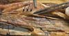
During a routine dissection of a formalin-fixed female cadaver utilized for educational purposes at the Faculty of Medicine of Aristotle University of Thessaloniki, the left SN was discovered presenting variant anatomical features and a distinct potential entrapment site. SN was formed by the anastomosis of the medial SN and the anastomotic peroneal branch of the lateral SN, arising from the tibial and common peroneal nerves, respectively. The anastomotic peroneal branch originated alone from the common peroneal nerve and did not give off any cutaneous branches, whereas the lateral cutaneous SN appeared absent. Moreover, the peroneal component of the SN was observed to take a course within a fibrous fascial tunnel 3.1 cm in length before it penetrated the crural fascia (Fig. 1). The tunnel was formed by a thickening of the fascia and was responsible for the nerve flattening and fixation that was enhanced by foot inversion. It should however be mentioned that the entrapment neuropathy diagnosis was not included in the patient's medical records and possibly overlooked.
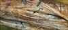
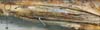
A second sensory nerve branch was observed originating from the common peroneal as a separate trunk lying underneath the crural fascia and medially to the anastomotic peroneal nerve of the SN (Fig. 2). The nerve took the place of the medial cutaneous branch, which typically arises from the lateral SN, being responsible for the sensory supply of the median and medial aspect of the calf. Between the middle and distal thirds of the leg, the supernumerary nerve crossed over the anastomotic peroneal branch and terminated before the formation of the SN, which occurred in the distal third of the leg (Fig. 3).
The cause of cadaver's death was unrelated to the present study, whereas neither evidence of previous surgical procedures undertaken at the region, nor pathological lesions at the area were encountered. The exposed anatomical structures were repeatedly documented by photography, while measurements were made by a metric electronic digital caliper (Mitutoyo Co., Tokyo, Japan) with an accuracy of 0.01 mm.
Discussion
The formation and topography of SN commonly present anatomical variability. A typical SN, formed by the medial SN and the anastomotic branch of the lateral SN, is found present in 40.2% [4] to 83.66% [5], while in 27.7% the nerve is formed at the distal third of the leg [4]. In the present case, the nerve is formed at the distal third of the leg (Fig. 3). Moreover, the anastomotic peroneal branch arose separately from the common peroneal nerve and participated in the distal formation of the nerve, although the typical anatomy of the lateral SN was not observed. Therefore, we could assume that in our case, the peroneal component of the SN was the lateral SN, which did not give off other cutaneous branches and became a real peroneal anastomotic nerve; this is noticed in 9.4% of the cases [4]. The anastomotic peroneal nerve has been mentioned to originate from the common peroneal nerve in a varying perchentage, ranging from 1.1% to 93% [4, 5]. During our dissection, another nerve was noticed taking proximally the place of the medial cutaneous branch of lateral SN. The lateral SN usually gives off branches which cross the medial SN at the middle of the leg in order to supply the medial surface of the calf [4], although in our case the noticed supernumerary nerve arose from the common peroneal nerve as a single trunk and lied medially to the peroneal component of the SN but laterally to the medial SN; it also crossed over the anastomotic peroneal branch again before its termination.
Entrapment of the SN could occur due to compression and fixation of the nerve caused by fascial thickening. Intrinsic causes, such as ganglions and lipomas [1, 3], as well as prolonged external compression by heel straps [2], positioning [6, 7], repetitive ankle sprains [2], fractures of the area [8] and gastrocnemius muscle injury [6, 7] may eventually lead to SN lesion. However, entrapment of the SN is mainly caused by compression and fixation of the nerve due to fascial thickening [3]. At the emergence point of the nerve, a fibrous arch is formed at the fascial opening through which SN passes, presenting a potential entrapment site. The "superficial sural aponeurosis" may be thickened and doubled, forming a fibrous tunnel that may compress the nerve [2]. In our specimen, the anastomotic peroneal branch of the SN traveled within a fibrous fascial tunnel that caused nerve flattening (Fig. 1). As we noticed, the tension was worsened during passive forcible plantarflexion and inversion of the foot, jeopardizing SN traction injury, as the nerve was tightly adherent to the surrounding fascia (Fig. 1). In the presented case, the flattening of the nerve within the fibrous tunnel indicates a potential entrapment site of the peroneal branch of the SN.
The diagnosis of entrapment neuropathy of the SN is based on clinical examination. The symptomatology includes sensory alterations over the distribution area of the nerve, thus the postero-lateral side of the distal third of the leg, as well as the lateral aspect of the foot and fifth toe. It should be mentioned that SN presents variable patterns of innervation over the dorsum of the foot, while it often communicates with the superficial peroneal nerve [9], leading to inconstant clinical findings after SN entrapment. Burning pain, hypaesthesia, dysaesthesia or paraesthesia, possibly with radiation over the foot or upper calf [2], may appear and worsen during night or exacerbate after exercise [1]; thereby dynamic examination may appear helpful [2]. In addition, tenderness may be observed over the nerve course, while pressure over the point of maximum tenderness can reproduce the symptoms [10].
In the present case, tenderness would be expected to appear over the entrapment site, thus in the lateral aspect of the calf and parallel to the course of the lateral SN, while the symptoms would be worsened during foot plantarflexion and inversion. The potentially non-typical appearance of the entrapment neuropathy may have leaded to misdiagnosis and justifies the report omission in the medical records. A positive Hoffman-Tinel's sign may affirm the diagnosis, although neurological examination could be normal. It is uncertain whether imaging techniques or electrodiagnostic studies can assist in establishing the diagnosis [2]. Doppler studies, ultrasonography, computed tomography or magnetic resonance imaging scanning may assist in visualization of the entrapment site, whereas increase in distal latency and decrease in amplitude of the sensory action potential of SN could indicate long-termed SN entrapment neuropathy [2, 6]. Moreover, injections with local anesthetic in the area of tenderness can lead to temporary resolution of the symptoms, representing an additional diagnostic tool [2]. We should furthermore highlight that SN entrapment neuropathy should be differentiated from other conditions, such as sacral 1 (S1) sciatica, exertional compartment syndrome, piriformis syndrome, popliteal artery entrapment that could present similar symptoms [2, 10].
Treatment of SN entrapment can be conservative or surgical. Non-operative approach may be initially preferred in cases of extrinsic etiology [3]. In these cases, avoiding or removing the offending agent may provide relief [3]. However unrarely, conservative treatment has been proved unsuccessful and surgical intervention is suggested [2]. Surgical removal of space-occupying masses is necessary, while in fascia-related cases of compression, neurolysis is the most effective technique [1]. In our case, SN decompression could have been accomplished under local anaesthesia, by a longitudinal section of the fascial tunnel that would release the nerve from the surrounding fibrous tissue. Knowledge of the etiology, symptomatology and treatment approach of such a condition is essential for surgeons and physicians, in order to establish a correct diagnosis and assist in improving patients' quality of life.
 XML Download