

 PDF
PDF ePub
ePub Citation
 Print
Print


Introduction
The inferior gluteal artery (IGA) gives three main branches in order as a ramus of articularis, a ramus of ischiadicus, and a ramus of muscularis during its extrapelvic course [1]. The IGA descends in the company of the sciatic nerve in the gluteal region, sending 2 to 4 muscular rami posterolaterally to gluteus maximus [2]. The distribution pattern of the IGA and the superior gluteal artery (SGA) has been reported in Korean [3]. The cadaveric study reveals that the IGA supplies a larger area of the gluteus maximus than the SGA did in most cases. But, the IGA is known to have minor contributions to the blood supply to the hip through an anastomosis with the medial circumflex femoral artery (MCFA) [4]. This report presents an occasional variation of the IGA observed on the buttocks during routine educational dissection course.
Case Report
During a routine dissection at Jeju National University Medical School in 2012, a case of an uncommon branching pattern with a prominent articular ramus of the IGA was observed in a 39-year-old Korean male cadaver, whose cause of death was rectal carcinoma. The protocol for the current report did not include any specific issue that needed to be approved by the ethics committee of our institution and it conformed to the provisions of the Declaration of Helsinki in 1995. A gross dissection was performed in the customary fashion. No other variations in the gluteal region were found during the dissection.
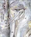
On the left gluteal region, the IGA gave muscular and articular branches at the inferior margin of superior gemellus during its extrapelvic course (Fig. 1). The IGA descends in the company of the sciatic nerve and the inferior gluteal nerve, sending 3 muscular rami posterolaterally to the gluteus maximus. The 3 muscular branches descend downwards in parallel with one another. The distances between the branches were 6 mm, 5 mm, and 4 mm from the medial aspect. Also the branches seemed to run downwards. An articular ramus crossed superficially to the sciatic nerve and continued medially running between the superior gemellus and the obturator internus. During its course, another muscular branch diverged from the articular branch. The articular branch travelled posterior across the obturator internus and the inferior gemellus and met the MCFA in the interval between the inferior gemellus and the quadratus femoris. The vessel formed from the anastomosis between the IGA and the MCFA is connected to the gluteus maximus muscle. Another muscular branch diverged from the articular ramus of the IGA and arose from the IGA at the inferior margin of the superior gemellus.
Discussion
The variations found in this case were as follows: The IGA branches off downwards at a time rather than in the usual order, the muscular rami ran in parallel with one another, and the articular ramus has an anastomosis with the MCFA.
The branching pattern and variation of the IGA and the SGA have been reported in 47 Korean adult cadavers [3]. According to the dissection study, the distribution patterns of the IGA and the SGA in the gluteus maximus were classified into four categories: I, the typical inferior type; II, the greater inferior type; III, the superior type; IV, the greater superior type. The variations found in this study correspond with the classification type I, which accounted for 47.3% of the cases. But, this case has uncommon branching patterns on its muscular rami as well. Interestingly, there is only a few case reports on the IGA variation that the IGA was entirely absent and the absence was compensated by a branch coming from the SGA [5, 6], which corresponds with the previous classification type IV. The classification of the distribution patterns provides useful information for flap surgery, because the variations of the arteries may make it difficult to find or separate those that are deep inside muscles [3]. On the aspect of practical application, the infragluteal perforator flap is known as a versatile flap for breast reconstruction [7] and for coverage of pressure sores of the sacrum and the perineal region [8].
In this case, the anastomosis between the MCFA and the IGA gives an interesting fact. In general, the IGA has minor contributions to the blood supply of the hip through an anastomosis with the MCFA adjacent to the tendon of the obturator externus [4]. But, recent reports revealed that the MCFA receives a direct supply from the IGA immediately before passing beneath the capsule of the hip [2, 8, 9]. In this case, the articular branch had a bigger external diameter than the muscular rami and gave another muscular branch during its course for an anastomosis with the MCFA. Although the direction of the blood flow in the anastomosed vessel is not clear from the post-mortem investigation, the characteristics of the articular branch were peculiar due to the cause of death, rectal carcinoma. As it is shown from this case and previous reports of the anastomosis between the MCFA and the IGA, the anastomosis between the MCFA and the IGA might be taught and regarded as normal, not rudimentary.
In conclusion, the branching pattern of the IGA was so distinctive that it may give useful information of versatile flaps for reconstruction and the prominent articular rami found in this case is good enough to confirm the existence of the anastomosis between the MCFA and the IGA.
 XML Download