

 PDF
PDF ePub
ePub Citation
 Print
Print


Introduction
The retromandibular vein (RMV) is formed within the parotid gland by union of the maxillary and superficial temporal veins. Within the gland, the RMV divides into anterior and posterior divisions. After exiting the gland, the anterior division joins the facial vein to form the common facial vein, which drains into the internal jugular vein; the posterior division of the RMV unites with the posterior auricular vein to form the external jugular vein (EJV). The EJV descends obliquely superficial to the sternocleidomastoid muscle and is accompanied by the great auricular nerve. The EJV then enters the roof of the posterior triangle of the neck and pierces the investing layer of deep cervical fascia to drain into the subclavian vein [1].
Subcutaneous veins of the head and neck show variations in their formation, topographic disposition, course, and draining pattern [2]. Detection of such anomalous venous patterns is essential for anatomists, clinicians, surgeons, and radiologists. Such anomalous venous patterns are relatively common in superficial head and neck veins. However, to the best of our knowledge, absence of the RMV in association with an atypical EJV and an anomalous vein has not been reported. We report such a case considering the possible consequences during various clinical procedures.
Case Report
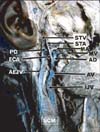
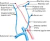
During routine cadaver dissection of the head and neck by medical undergraduate students, we observed a rare venous variation in the right neck of a male cadaver aged about 55 years at the time of death. The RMV was absent unilaterally. The maxillary vein was bifurcated into anterior and posterior divisions (Fig. 1). The posterior division united with the superficial temporal vein to form an atypical EJV in the parotid region, which descended obliquely, superficial to the sternocleidomastoid muscle. The atypical EJV was accompanied by the great auricular nerve and drained into the subclavian vein. The anterior division of the maxillary vein joined the facial vein to form an anomalous vein in the submandibular region (Fig. 2). The anonymous vein descended deep to the stylohyoid and posterior belly of the digastric muscle, crossed superficial to the external carotid artery, and drained into the internal jugular vein.
Discussion
Head and neck veins exhibiting variations or malformations are important during clinical applications such as reconstructive microsurgeries and diagnostic procedures. Therefore, knowledge of the possible anatomical variants of these veins helps to avoid misinterpretations or misidentifications during conventional radiographic procedures such as angiography and venous catheterization [3].
Among the superficial veins of the head and neck, the EJV often shows variation both in its pattern of origin and termination. Because of its known and frequent variability, it is likely improbable to determine the normal pattern of this vein [4].
Though variations in superficial veins of the head and neck are uncommon, the RMV is exceptional, with some authors reporting that it may show variations in its division pattern [2, 3]. However, to the best of our knowledge, no study has reported its total absence.
Venous patterns of the head and neck develop immediately following skull development. Venous pattern development is a complex process initiated by formation and eventual regression of the cephalic veins and is associated with formation of interconnecting venous spaces, giving the appearance of a plexiform arrangement. Upon further development, selective "retention" and "regression" of some network channels results in a definitive venous pattern [5]. Thus, the EJV and RMV develop as secondary channels from a capillary plexus derived from a tributary of the cephalic vein in tissues of the neck and temporal region, respectively [6]. Developmental errors during crucial stages of venous formation result in an abnormal venous pattern or the absence of a particular venous channel.
In cases of an undivided RMV, it most often continues as the EJV [2, 7, 8], or it may join the facial vein to form the common facial vein [3]. An undivided RMV is found in 1 out of 104 cases [9]. When the RMV is undivided and there is no common facial vein, the RMV may join with the internal jugular vein and interfere with blood drainage from the anterior face.
Variant patterns of superficial head and neck veins are vulnerable to profuse bleeding during neck surgery. Hence, it is advisable to have knowledge of an abnormality before performing any clinical or radiological procedures in this region.
The RMV is used as a guide to expose the branches of the facial nerve during superficial parotidectomy and open reduction of mandibular condylar fractures [10]. In these procedures, using the RMV as a guide to expose the branches of the facial nerve is identical to the procedure for exposing the condyle and elevating the superficial lobe of parotid gland, thus minimizing surgical stress [11].
The superficial course of the EJV is easier to follow with a venous manometer as it is more readily accessible than the internal jugular vein. The EJV is also used in catheterization for hemodialysis as it involves a simple, short procedure without any severe complications [12]. An abnormal pattern of the EJV, particularly variant formation and termination, may interfere with this approach.
In conclusion, variant patterns of venous drainage from the head and neck region are uncommon. This is the first report showing complete absence of an RMV with abnormal division of the maxillary vein and formation of an atypical EJV.
 XML Download