

 PDF
PDF ePub
ePub Citation
 Print
Print


Introduction
The gallbladder is the main part of the extrahepatic biliary system. Double gallbladder is a rare congenital anomaly occurring in 1/4,000 births [1, 2]. Failure to detect this anomaly is problematic for diagnosis and treatment of cholecystitis, which might result in recurrent cholecystitis postoperatively [3]. Therefore, preoperative diagnosis is essential, as it is difficult to locate the accessory gallbladder during surgery [4]. Double gallbladder affects males and females relatively equally; however, owing to a higher incidence of gallbladder disease in females, the number of cases of double gallbladder is higher in females than in males [5].
A cystogastric fold is a peritoneal fold that extends from the lesser curvature of the stomach to the gallbladder. Peritoneal folds extending from the stomach and duodenum to the gallbladder are very rare. To the best of our knowledge, few reports are available discussing folds connecting the stomach, lesser omentum, and greater omentum with the gallbladder [6, 7]. Thus, we report a rare case of double gallbladder where both the main and accessory gallbladders were enclosed in a cystogastric fold. We hope this report alerts radiologists and laparoscopic surgeons to the possibility of a double gallbladder in a cystogastric fold, as these variations might result in misinterpretations, misdiagnosis, and surgical or postoperative complications.
Case Report
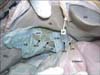
During dissection classes for medical undergraduate students, we observed variations in the gallbladder of a 70-year-old adult male cadaver. A cystogastric fold extended from the lesser curvature of the stomach and the first part of the duodenum to the gallbladder, and the left end of the fold merged with the lesser omentum (Fig. 1). The fold enclosed two gallbladders: the main gallbladder and the accessory gallbladder. The larger of the two gallbladders was dumbbell shaped, and was 9 cm long and 3 cm at its broadest part (Fig. 2). The fundus was attached to the anterior part of the gallbladder fossa. The body was within the cystogastric fold. The main cystic duct arose from its neck and was straight. It measured 25 mm in length and 5 mm in breadth. The accessory gallbladder was a small pouch 2 cm long and 1.5 cm broad. It was situated in the posterior part of the gallbladder fossa and was separated from the main gallbladder, as the body and neck of main gallbladder were suspended in the cystogastric fold. The fundus was attached to the main gallbladder by fibrous tissue, while its duct joined the distal part of the main cystic duct (Fig. 2). The lumen of the accessory gallbladder had green bile residue, and its mucosa had typical folds as seen in the main gallbladder. The duct from the accessory gallbladder was 2 cm in length. The fundus of the main gallbladder did not project beyond the cystic notch. The accessory gallbladder was supplied by the intrahepatic branches of the right hepatic artery. There was no additional cystic artery passing to the accessory gallbladder through the Calot's triangle.
Discussion
Double gallbladder is a rare congenital anomaly. According to Boyden [2], abnormal differentiation of the primordial gallbladder during the fourth and fifth week of gestation might result in formation of multiple gallbladders. Gallbladder duplication is classified into two major groups by Boyden [2] based on the presence or absence of a common cystic duct. In the first group, the duplicated gallbladder shares the same common cystic duct, while in the second group, the gallbladder duplication is associated with a separate cystic duct for each gallbladder. The current case belongs to the first group where there two cystic ducts join to form a common cystic duct before joining the common hepatic duct. Cases of double gallbladder have been reported in the past. They have been observed in cadaveric dissections, autopsies, radiological examinations, and surgeries. Sangeeta et al. [8] reported the presence of double gallbladder from cadaveric dissections. Abdominal ultrasonography is a technique that can reveal the presence of double gallbladder. Vijayaraghavan and Belagavi [9] and Pasha [10] reported cases of double gallbladder detected using ultrasonography. In spite of radiologic examination, cases of double gallbladder may be missed during radiological interpretation and even during surgery. Such cases will be evident postoperatively when symptoms occur.
The gallbladder is rarely enclosed partly or completely in a peritoneal fold [6, 7]. Such a fold might result in reduced size or function. It might also lead to problems in diagnosis and laparoscopic surgery. The peritoneal fold that enclosed the gallbladder in this case did not compress the gallbladder, which was evident by the size of the gallbladder. This fold was likely derived from the ventral mesogastrium since the gallbladder, along with the liver, develops in the ventral mesogastrium, and its continuity with the lesser omentum is suggestive of the same. Presence of double gallbladder and a cystogastric fold is extremely rare and, to the best of our knowledge, this is the first report of such a case. Awareness of this anomaly will help radiologists avoid misinterpreting images and help surgeons reduce complications following laparoscopic surgery.
In conclusion, the awareness of the presence of a double gallbladder in a cystogastric fold may improve the success of laparoscopic cholecystectomy and liver surgeries, and help in the interpretation of cholecystography findings.

 XML Download