

 PDF
PDF ePub
ePub Citation
 Print
Print


Introduction
Uncontrolled self-renewal is proposed to be an important mechanism in carcinogenesis [1]. On the basis of the cancer stem cell (CSC) hypothesis, a tumor may be sustained by a subset of cancer cells with stem cell-like features that have the ability for self-renewal and pluripotency [2, 3]. These CSCs have tumorigenic potential and proliferate indistinctly [4]. The same molecular pathway that manages self-renewal in normal stem cells also seems to manage CSCs in tumors [5]. Octamer 3/4 (Oct 3/4), a member of the POU family, is considered to be an important stem cell marker and essential transcription factor during human embryogenesis [6]. In recent years, there have also been reports about the presence of Oct 3/4 in differentiated benign and malignant human cells [7]. Sox2 is a member of the Sox (SRY-Related HMG box) gene family that encodes transcription factors with a single HMG DNA-binding domain. Sox2 controls neural progenitor cells by prohibiting their ability to differentiate [8]. Sox2 is also expressed in several malignant tissues [9]. Nanog is believed to function in conjunction with other factors such as Oct4 and Sox2 to form an embryonic stem cell identity [10, 11]. Recently, Nanog expression has been reported in human neoplasms, including germ cell tumors, breast carcinoma, and osteosarcoma [12]. Nucleostemin is expressed in the nucleoli of adult central nervous system stem cells, primitive bone marrow cells, embryonic stem cells, and several cancer cell lines [13]. Nucleostemin is often used as a stem cell marker [14]. Bmi is a potent repressor of retinoblastoma and the p53 pathway, its role in tumorigenesis is very important [15]. It is a transcriptional repressor that belongs to the polycomb-group family of proteins that are involved in hematopoiesis, regulation of proliferation, and senescence [16]. The Bmi gene is widely expressed in diverse human tumors including lymphomas, bladder and breast carcinomas, and neuroblastoma [17]. Zfx is a zinc finger protein of the Zfy family. Zfx normally suppresses apoptosis in embryonic stem cells. Zfx directly activates common target genes in embryonic and hematopoietic stem cells, as well as embryonic stem cell-specific target genes such as embryonic stem cell self-renewal regulation of Tbx3 and Tcl1 [18]. The Tbx3 gene shares a common DNA binding domain. The T-box genes encode transcription factors involved in regulation of the developmental process [19, 20]. Tcl1 has been extensively studied as an oncogene in T-cell leukemia and plays an important role in early mouse embryos and embryonic stem cells [21]. Esrrb in coordination with Nanog and Oct4 activate the internal machinery of ES cells [22]. The Dppa4 protein is located in the embryonic stem cell nucleus and is associated with chromatin. It regulates the differentiation of ES cells into a primitive ectoderm lineage [23]. Although the expression of these genes in embryonic stem cells has been extensively studied, little is known about their expressions in somatic cancers. Colon, prostate, bladder, and hepatocarcinomas are common worldwide carcinomas, whose diagnoses and treatments are challenging [24, 25, 26]. We have examined the expressions of Oct4, Sox2, Nanog, nucleostemin, Bmi, Zfx, Tcl1, Tbx3, Dppa4, and Esrrb these genes in various cancers. It is possible that these genes participate in the development and regulation of tumor and hyperplastic tissues. Thus, we have used a number of malignant tumor tissues and cancer cell lines to investigate the expressions of these genes with the intent to study the mechanism of cancer cell self-renewal.
Materials and Methods
Sample collection
Prostate, bladder, and colon cancer samples were obtained from patients who referred to Tohid Hospital, Sanandaj and Imam Khomeini Hospital, Tehran, Iran. Normal tissue samples (apparently normal tissues from the margins of colon cancer samples) were collected. Samples were immediately snap-frozen in liquid nitrogen and stored at -70℃ until used for RNA extraction. All patients consented to have their biopsy samples analyzed. This study was approved by the Ethics Committees of Kurdistan University of Medical Sciences.
Cell lines and cultures
LNCaP (human prostate cancer), HepG2 (hepatocarcinoma), HT-1376 (bladder cancer), U87 (glioblastoma), NT2 (embryonal carcinoma stem cell), HT-29, and Caco-2 (colon cancers) were maintained at 37℃ in a humidified atmosphere of 5% CO2 with RPMI1640 medium (Gibco, Paisley, UK), supplemented with 100 U/ml penicillin, 100 µg/ml streptomycin, and 10% fetal bovine serum (FBS; Gibco). Caco-2 and NT2 were cultured in Dulbecco's modified Eagle medium supplemented with 15% FBS. Passaging was routinely performed with 0.25% Trypsin/EDTA.
RNA extraction and reverse transcriptase polymerase chain reaction
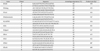
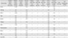
Total RNA was isolated from frozen tissues and cell lines using the RNX-Plus solution (CinnaGen, Tehran, Iran) according to the manufacturer's instructions. Purity and integrity of the extracted RNA was measured by UV spectrophotometry (260/280 nm ratio) as well as visual observation of samples by agarose gel electrophoresis. Total RNA was treated with RNase-free DNase (Fermentas, St. Leon-Rot, Germany) for which 1 µg was used for cDNA synthesis, with the RT Kit (reverse transcriptase polymerase chain reaction [RT-PCR] Kit, Bioneer, Daejeon, Korea) and random hexamer primer in a 20 µl reaction according to the manufacturer's instructions. The RT-PCR Kit consisted of 5× buffer, dNTP, RNAsin and RT enzyme (RevertAid M-MuLV reverse transcriptase). For each sample, a non-RT control was used in parallel to detect any potential nonspecific ampli fication of contaminated genomic DNA. PCR primers were designed by Gene Runner and Prime 3 software. The primer sequences of the genes are shown in Table 1. PCR was conducted with 2 µl of cDNA sample, 0.2 µl of Taq polymerase (CinnaGen), 0.75 µl MgCl2 (25 mM), 0.5 µl dNTP (10 mM), 2.5 µl 10× buffer, 0.5 µl of each primer (20 mM), and 18 µl dH2O. We used glyceraldehyde 3-phosphate dehydrogenase (GAPDH) as an internal standard for RT-PCR. PCR amplification was performed for either 36 (Oct4, Nanog, Sox2, Bmi), 40 (nucleostemin, Tcl1, Esrrb, Dppa4), or 32 (Tbx3, Zfx) cycles with the following cycle conditions: 94℃ for 45 seconds, 60℃ (Oct4, Nanog, Sox2, nucleostemin); 59℃ (GAPDH); 58℃ (Tbx3); and 62℃ (Dppa4, Esrrb) for 45 seconds by the Thermal cycler (Eppendorf, Hamburg, Germany). PCR products were separated on a 1% agarose gel, stained with ethidium bromide and then visualized and photographed with a UV trans-illuminator (UVIdoc, Cambridge, UK).
Immunocytochemistry
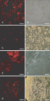
The expressions of Oct4 and nucleostemin at the protein level were determined by immunostaining in the LNCaP, HepG2, and HT-1376 cell lines.
Briefly, different cell cultures were plated in 4-well dishes until confluent. Next, cells were fixed with 4% paraformaldehyde for 15 minutes at room temperature. After washing with phosphate buffered saline (PBS), fixed cells were permeablized with 0.25% Triton X-100. The cells were then incubated in the appropriate diluted (1:50) first anti bodies (affinity-purified goat polyclonal anti-Oct4 for Oct4; and anti-nucleostemin for nucleostemin; Santa Cruz Biotechnology) in PBS that contained 1% bovine serum albumin (BSA) for 1 hour at room temperature. After washing for three times with PBS, cells were incubated with the appropriate secondary antibodies (Texas red-conjugated donkey anti-goat for Oct 4 or FITC-conjugated donkey anti-goat for nucleostemin, Santa Cruz Biotechnology, Santa Cruz, CA, USA) at a dilution of 1:100 in 1% BSA for 1 hour at room temperature. Cells were then washed three times with PBS. For the negative control all conditions were kept the same, except that the primary antibody was omitted. Fluorescent images were obtained with a Nikon TE-200 fluorescent microscope equipped with a digital camera (Nikon, Tokyo, Japan).
Results
Expression of stem cell markers in cancer tumor tissue specimens and in cell lines
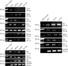
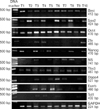
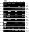
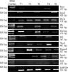
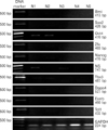
Stem cell markers were expressed in malignant human colon, prostate and bladder tissues as well as the cancer cell lines (HT-29, Caco-2, HT-1376, LNCaP, and HepG2). Specific primers were used to amplify segments of the Oct4, Nanog, Sox2, nucleostemin, Bmi, Zfx, Esrrb, Dppa4, Tcl1, and Tbx3 genes and examined their expressions by RT-PCR (Table 1). As expected, DNA fragments of these genes, based on the designated primers (Table 1), were amplified in the PCR reactions.
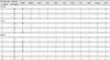
Specific primers for Oct4 have been designed [27] to amplify a particular segment of Oct4 that is common in both spliced variants of the gene (GenBank accession numbers: NM_002701 and NM_203289). We detected the expression of Oct4 in all (100%) tumor samples (colon, bladder, and prostate) as well as the hepatic cancer cell lines (Tables 2, 3, Figs. 1, 2, 3, 4, 5, 6). Nanog expression was detected in 100% of the colon cancer samples, 90% of the bladder cancer samples, and 80% of the prostate cancer samples. Nucleostemin was detected in 100% of the prostate cancer samples, 80% of the bladder cancer samples, and 60% of the colon cancer samples (Tables 2, 3, Figs. 1, 2, 3, 4, 5, 6).
As seen in Figs. 1, 2, 3, 4, 5, 6, 7 and Table 2, expressions of all ten genes were detected in 40% to 100% of the bladder cancer tissue, and 60% to 100% of both colon and prostate tumor samples.
We also checked for expression of the ten genes in five normal colon tissues. Only low-level expressions of Oct4 and NS genes were detected (Fig. 5).
There was a significant relation between tumor grade and expression of self-renewal genes. In the prostate cancer samples, 60% of the self-renewal genes were expressed in grade I, 80% in grade II, and 90% in grade III tumors. In colon cancer samples, 40% to 100% of the self-renewal genes were expressed in grade I, 70% in grade II, and 60% to 80% in grade III tumors. Bladder cancer samples expressed about 60% of the self-renewal genes in grade I tumors, 70%, in grade II tumors, and 100% in grade III tumors. Expression of Oct4 was detected in all tumor grades of the prostate, colon, and bladder cancer samples. Expressions of Oct4, Nanog, Sox2, NS, Zfx, Tbx3, Tcl1, and Esrrb genes were detected in 100% of the grade III cancer samples from prostate, colon, and bladder tissues (Table 3, Fig. 6).
Immunocytochemistry
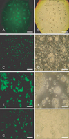
We used the polyclonal anti-Oct4 and anti-nucleostemin antibody to examine subcellular localizations of Oct4 and Nucleostemin proteins in the cancer cell lines by immunocytochemistry (ICC). NT2, a human embryonic carcinoma cell line, was the positive control, which showed the same pat tern of antigen expression as previously described [28]. U87, a human glioblastoma cell line, was used as the positive control to optimize the experiment for NS, which has nucleolar localization for nucleostemin. Our results confirmed expression of Oct4 (Fig. 7) and nucleostemin (Fig. 8) in the cancer cell lines. There was no staining signal in the negative controls, where all conditions were the same with the exception of the primary antibodies. The experiment was repeated twice, with similar results.
Discussion
A specific aim in cancer research is to determine the mecha nism by which CSCs arise and self-renew [1, 2, 29]. The CSC hypothesis proposes that only a small degree of tumor cells are enriched for cancer initiation, causing tu mor development [1, 2, 3]. This new idea of cancer may also alter our diagnostic and therapeutic goals by allowing us to better determine and target CSCs [5, 6]. Uncontrolled self-renewal plays a direct function in different types of carcinoma progression, or profits as a precious marker of tumorigenesis [4, 5, 29].
In this study, we have examined the expressions of self-renewal regulatory factors such as Oct4, Nanog, Sox2, nucleostemin, Bmi, Zfx, Esrrb, Tcl1, Tbx3, and Dppa4 in different cancer types and cancer cell lines. These genes are required for efficient self-renewal of embryonic stem cells [12, 13, 14, 15, 16, 17].
In many germ cell tumors as well as a few somatic tumors, there is observable expression of Oct4 [9, 30, 31]. Oct4 can function as a potent oncogene in vivo and support the image that progenitor cells are an energetic force in tumorigenesis [32]. Oct4 expression has been detected in human breast, liver, pancreas, colon, kidney, and gastric cancers, as well as in Hela and MCF7 cells, and bladder tumors [33, 34, 35, 36, 37, 38].
In this study, we detected the expression of Oct4 in all cancer cell lines. ICC analysis further determined the expression of Oct4 at the protein level. Looijenga et al. [35] have previously reported a lack of expression of Oct4 in a panel of somatic tissues such as bladder tumors [35], this finding does not agree with our study conclusion. Possibly, this inconsistency might be due to the heterogeneous nature of tumors [36]. Recently, several groups have reported the expression of Oct4 protein in several somatic tissues [37, 38]. In 2007, Atlasi et al. [33] have reported expression of Oct4 in bladder tumors, which confirmed our results. In our study, Nanog expression was detected in 100% of the colon cancer samples, 90% of the bladder cancer samples and 80% of the prostate cancer samples. Recently, Nanog expression has been reported in human neoplasms, including germ cell tumors, breast carcinoma and osteosarcoma [12, 13]. Ezeh et al. [12], in 2005, have reported elevated expression levels of Oct4 together with Nanog, Stellar, and Gdf3 mRNA in both breast carcinoma tissue and breast carcinoma cell line MCF7. These results have suggested that breast carcinomas, and possibly other human malignancies, may contain cells remnant of embryonic-like stem cells [11, 12]. In addition, a clinical survey showed that elevated expression of Nanog was related to retinoblastoma, prostate cancer, embryonal carcinoma, metastatic germ cell tumor, and ovarian cancer [38]. Taken together, these findings have indicated that abnormal expressions of Oct4 and Nanog in stem cell and tumor tissues might play a vital role in tumor transformation, tumorigenicity, and tumor metastasis [39, 40]. Hoei-Hansen et al. [38] has stated that Sox2 is overexpressed in patients with ovarian carcinoma. In the present study, we have found the expressions of Oct4, Nanog, and Sox2 in several human tumors. In addition, we observed low expression of Oct4 in some normal colon tissues that were obtained from asymptomatic individuals. Recently, Tai et al. [7] have shown that Oct4 is expressed in several adult human stem cells and Matthai et al. [37] have reported Oct4 expression in normal human endometrium. The observed expression of Oct4 in normal colon tissues might reflect the presence of rare normal colon stem cells in these samples.
Nucleostemin expressed in human placental tissues also at a high level, possibly due to the existence of a mass of placental stem cells [40, 41]. In the present study, nucleostemin highly expressed in cancer tissues and cancer cell lines. We detected poor expression of nucleostemin in some normal colon tissues. Recently, a small quantity of nucleostemin expression has been detected in normal human muscle [42, 43], possibly in few myoblasts with stem cell characteristics existing in muscle tissues [43]. ICC demonstrated that LNCaP, HepG2, and HT-1376 cancer cell lines showed cellular staining for nucleostemin proteins. These results were similar to those of previous studies of other human cancers. In normal stem cells and U87-glioblastoma cell line, nucleostemin protein has been shown to localize in the nucleolus [13, 44]. However, cancer cell lines that we used exhibited both cytoplasmic and nucleolar localizations of nucleostemin. This distribution pattern of nucleostemin has recently been observed in gastric, prostate, and renal cell carcinomas [44, 45]. Recently, a novel nucleostemin binding protein was identified as an alpha isoform of human protein phosphatase 2 regulatory subunit B (B56 PPP2R5A). PPP2R5A is located in the cytoplasm [46], which may explain to some extent why nucleostemin protein displays both cytoplasmic and nucleolar locations; however, further investigations are necessary to determine why it occurs at this exact location.
In this study, Bmi overexpressed in human bladder, colon and prostate cancers as well as cancer cell lines. The results of our research were consistent with previous reports from other cancers. The Bmi gene is widely expressed in diverse human tumors such as lymphoma, bladder cancer, breast cancer, and neuroblastoma [16, 17, 18]. There are numerous reports on the expressions of Zfx, Tcl1, Tbx3, Esrrb, and Dppa4 in cancers. In this study, we have aimed to explore the expressions of these genes in human cancers. RT-PCR analysis confirmed expressions of Zfx, Tcl1, and Tbx3 in human bladder, colon and prostate cancers as well as cancer cell lines. Zfx is a transcription factor specifically required in ESC embryonic and adult stem cell populations such as hematopoietic stem cells. In addition to common target genes [20], the Zfx gene is highly expressed in glioma tissue and cell lines [47]. In addition, it can directly activate cell type-specific targets such as Tbx3 and Tcl1 in embryonic embryonic stem cells [21]. The pattern of Tcl1 expression as well as its defects from hematopoietic stem cells indicates its potential to act as a highly specific drug target in specific malignancies of lymphoid and germ-cell origin [22]. Lau et al. [48], in 2010, have shown protein expression of Tcl1 in testicular germ cell tumors. Overexpression of Tbx3 has been described in breast, liver, and ovarian cancers in addition to endometrioid adenocarcinomas [49, 50, 51]. Based on the key role of Tbx3 in carcinogenesis, inhibition of Tbx3 activity could be an effective therapeutic strategy for human cancer [52]. Esrrb has been reported to interplay with the ES cell embryonic stem cell master regulator Nanog. Many of these potential Esrrb target genes comprise Oct4, Sox2, and Nanog-regulated genes and include the respective promoters of these core ES cell embryonic stem cellregulators [53]. Expression of the Dppa4 gene in embryonic stem cells has been studied, but little is known about its expression in somatic cancer [23]. There is no published report on the expression of these genes in cancers. We reported the expressions of Dppa4, Esrrb, Tcl1, Tbx3, and Zfx in human bladder, colon and prostate cancers as well as cancer cell lines. The results of our research confirmed previous reports of other cancers.
In conclusion, our data is a report on the expression of the ES cell marker Nanog, nucleostemin, Sox2, Zfx, Tcl1, Tbx3, Esrrb, and Dppa4 in bladder, prostate, and colon cancers as well as in cancer cell lines. Therefore, this data might provide valuable information on the nature and behavior of tumors, leading to a new strategy for targeting CSCs and perhaps one step closer to the prevention of cancer recurrence and metastasis. However, further studies are required to isolate and characterize the putative CSCs from tumors and elucidate the role of gene self-renewal in tumor carcinogenesis.

 XML Download