

 PDF
PDF ePub
ePub Citation
 Print
Print


Introduction
The liver is the largest gland in the human body. It is situated below the right dome of the diaphragm and mainly occupies the right hypochondriac, right lateral/lumbar, and epigastric regions of the abdominal cavity. It is divided into anatomical right and left lobes by the reflection of the falciform ligament on the anterior surface, the fissure for ligamentum teres on the inferior surface, and the fissure for the ligamentum venousum on the posterior surface. The anatomical right lobe is larger than the left lobe and has caudate and quadrates lobes as parts. The quadrate lobe is situated between the fissure for the ligamentum teres and the fossa for the gall bladder, whereas the caudate lobe is situated between the fissure for the ligamentum venosum and the groove for the inferior vena cava. The gall bladder fossa is situated to the right of caudate lobe and contains the gall bladder. The gall bladder fundus projects beyond the inferior border of the liver and touches the anterior abdominal wall. A small, shallow notch is seen on the inferior border near the fundus. An area called the porta hepatis is on the inferior surface of the liver and allows the passage of blood vessels into the liver. The right and left hepatic ducts leave the liver through the porta hepatis and join to form the common hepatic duct. The cystic duct joins the common hepatic duct to form the bile duct just below the porta hepatis. Knowledge of lobe and fissure variations in the liver is of immense clinical and surgical importance [1]. The absence of normal fissures and lobes or the presence of additional lobes and fissures might lead to confusion on a radiological diagnosis of a liver disorder [2]. We observed variations in the external appearance of a liver and formation of the main duct system below the liver. We feel that this may be of interest to surgeons and radiologists.
Case Report
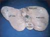
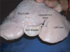
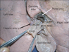
We found an anatomically peculiar liver during a routine dissection class for undergraduate medical students. Th is liver was found in an adult male cadaver aged approximately 60 years. The quadrate lobe and the fissure for the ligamentum teres were totally absent in this liver (Fig. 1). The cystic notch on the inferior border was very broad and was 5 cm deep. Thus, the fundus and body of the gall bladder projected out onto the anterior surface of the liver (Fig. 2). The ligamentum teres appeared to enter through the anterior surface of the liver because of the depth of the fissure, which was just to the left of the gall bladder. The cystic duct terminated into the right hepatic duct at the porta hepatis instead of opening into the common hepatic duct (Fig. 3).
Discussion
The liver is known to show lobe and fissure anomalies, and knowledge of these anomalies is useful to the radiologist when interpreting liver radiologic findings. The presence of additional lobes and fissures or the absence of normal lobes and fissures might lead to confusion during surgery or clinical misdiagnosis. Such clinical misdiagnoses have occurred in the past when there was an additional lobe [2]. Several lobe and fissure anomalies of the liver have been noted and reported in the past. Joshi et al. [1] reported notching along the inferior border of the caudate lobe in 18% of livers, a vertical fissure in 30%, and prominent papillary process in 32% of livers in their extensive study on lobes and fissures of the liver.
Fitzgerald et al. [2] reported the presence of an additional lobe. Preoperative imaging of this lobe led to the misdiagnosis as a lesser omental lymphadenopathy. The presence of a large symptomatic accessory liver lobe has been reported in a woman aged 70 years [3]. Pujari and Deodhare also reported the presence of a symptomatic accessory lobe. Accessory lobes may herniate into the thorax through the diaphragm and can cause serious problems. A case of a bifid liver presenting with anomalous quadrate and caudate lobes and a transverse gallbladder has been reported recently [4]. A few cases of hypoplasia of the left lobe of the liver have also been reported. Very recently, Anjamrooz and Azari [5] reported a case of the coexistence of multiple anomalies of the hepatobiliary system. Reports on the presence of accessory liver sulci are also available [6]. However, it is very rare to have the total absence of any of the liver lobes. In the current case, the quadrate lobe was totally absent; thus, the gall bladder fossa was broad and there was an associated absence of the fissure for the ligamentum teres. When the quadrate lobe is absent, the gall bladder is shifted more to the left , which could be important for surgeons performing laparoscopic cholecystectomy.
The gall bladder and cystic notch can also show variations. A few cases of a left sided gall bladder and right sided ligamentum teres have been reported [7, 8]. The gall bladder may be short or atrophic when abnormal peritoneal folds such as cystohepatocolic folds are present [9, 10]. Short gall bladders that hide in their fossa may lead to confusion during imaging and laparoscopic surgery. In the current case, the cystic notch was too deep and wide. Thus, the fundus and body of the gall bladder were projecting between the right and left lobes of the liver giving a peculiar look to the liver. This might make the gall bladder vulnerable to blunt trauma, as a major part of the gall bladder touches the anterior abdominal wall. The deep cystic notch and exposure of the gall bladder between the right and left lobes of the liver might make the radiologist think of a congenital anomaly or a traumatic laceration of the liver that has forced the gall bladder to the front.
In the current case, the cystic duct opened into the right hepatic duct instead of opening into the common hepatic duct. The termination of the duct was at a much higher level than the normal termination of the cystic duct. This might not have lead to any functional disturbance but might give a very small space for the surgeon to ligate the cystic artery or even lead to confusion finding it.
We conclude that knowledge of the absence of a quadrate lobe and the presence of a deep cystic notch is important for planning a laparoscopic cholecystectomy. The projection of gall bladder on the anterior surface of liver can make it vulnerable to blunt injury of the upper abdomen.
 XML Download