

 PDF
PDF ePub
ePub Citation
 Print
Print


Introduction
Congenital anomalies of the rotation and fixation of the gut are common in children but exceedingly rare in adults [1]. Textbooks of anatomy describe the sigmoid colon as viscera having a constant morphology with variations in its length. Other authors have classified the sigmoid colon into several types according to its position and morphology. The present study reports a morphologically abnormal descending and sigmoid colon with its clinical implications and embryological basis that has not been reported earlier. Knowledge of the variation in morphology and position of the sigmoid colon is of value to surgeons and radiologists for interventional procedures, preventing misdiagnosis of an abnormally positioned sigmoid colon.
Case Report
Methods
During a routine cadaveric dissection at the Department of Anatomy, Lady Hardinge Medical College and SSK Hospital, New Delhi, India, the abdomen of a 54-year-old man of Indian origin showed a right-sided descending colon and sigmoid colon loop with a common mesentery. The colon was carefully dissected. The morphology was studied in detail with special reference to its position, attachment of sigmoid mesocolon, and location of other viscera close to the sigmoid colon.
Results
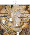
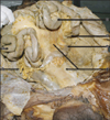
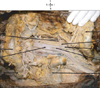
For convenience of description, the anomalous colon was divided into four parts (Fig. 1). The first part stretched obliquely from the splenic flexure to the right side along the root of the mesentery, traversing the umbilical quadrant, 3.5 cm to the right of midline at the level of L5. The second part turned upward and toward the right, ascending up to the level of L2. The third part descended obliquely on the right side of the ascending limb up to the pelvic brim. The fourth part was in the lesser pelvis extending from the right sacroiliac joint to third sacral body (Fig. 2).
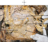
The first part of the colon with its oblique course was related posteriorly to the anterior surface of left kidney and suprarenal gland (obliquely from left to right), gonadal vessels, and lateral end of renal vessels. It continued to descend obliquely and crossed the midline in front of the bifurcation of the aorta. On the right side just above the pelvic brim, it was related posteriorly to the right common iliac vessels, ureter, gonadal vessels, and psoas major. The cecum, appendix, and ileocecal junction were posterior to the second part of the colon in the right lumbar region, 3.5 cm above the transtubercular plane (Fig. 3). The third part was in the right iliac fossa related to the psoas major and iliacus muscle, posteriorly. The fourth part was anterior to the right ureter, and external and internal iliac vessels, at the pelvic brim. Within the lesser pelvis, the fourth part was related to the piriformis and superior rectal artery posteriorly.
Vascular supply
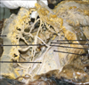
The inferior mesenteric artery (IMA) supplied this anomalous colon and had an unusual course. It was found to arise 3.5 cm below the lower border of the horizontal part of duodenum. In front of the aortic bifurcation, the IMA turned to the right instead of the left, crossing the right common iliac artery, lying medial to the right ureter, with the inferior mesenteric vein on its left (Fig. 4). It gave off the following three branches: The first branch (left colic artery) supplied the proximal segment of the first part of the anomalous colon forming a marginal artery. The distal segment of the first part was supplied by the second branch of the IMA (superior sigmoid artery). The second, third, and fourth parts of the colon were supplied by the inferior sigmoid branch of the IMA, which arose from the right side of the IMA instead of the left (Fig. 5).
Discussion
Parts of the anomalous colon were differentiated by their arterial supply [2]. The second, third, and fourth parts were supplied by one artery, whereas the first part was supplied by two arteries. The proximal segment of first part of the colon can be delineated as the descending colon, as it was supplied by branches of the left colic artery, forming the marginal artery. The distal segment of the first part of the colon was supplied by the superior sigmoid artery, hence designated as the proximal part of sigmoid colon. The second, third, and fourth parts of the colon were supplied by branches of the inferior sigmoid artery. During surgery, the blood supply can be used to identify the segments of the colon. The sigmoid colon thus had an oblique proximal segment (left to right), a loop as the middle segment, and another oblique segment (right to left) that entered the lesser pelvis from the right side at the right sacroiliac joint. This type of sigmoid colon appeared to have a bent hairpin shape ( ).
).
).According to Madiba and Haffajee [3], the present case falls into the primitive variety, suprapelvic in position but on the right side. In this present case, the right sigmoid loop lay anterior to the cecum in contrast to the case described by Madiba and Haffajee [3]. It can be classified as type 5 in the detailed classification of Madiba and Haffajee [3], which has not been reported before.
Madiba and Haffajee [4] had proposed another way of classifying the sigmoid colon according to its level of origin from the descending colon. These were high, intermediate, and low. The present case is of intermediate variety since the descending colon was short and mobile immediately after the splenic flexure.
The right sigmoid colon has been reported radiographically with barium enema, by Fiorella and Donnelly [5] and Saxena et al. [6] in American and Indian children, respectively, <5 years of age. Komiyama and Shimada [2] have also described a right-sided sigmoid loop behind the ascending colon in an adult. Considering these reports, the present case stands as a rare finding in adults in describing an intermediate origin of the sigmoid colon with a bent hairpin shape lying on the right side anterior to the cecum and ascending colon.
Embryological basis
The primary ontogenic factors responsible for positional variations of the colon are the differential development of abdominal organs with their peritoneal coverings and a mechanical factor [7]. The reduction of physiological hernia starts with the cranial limb of the herniated gut passing to the right of the caudal limb and the intra-abdominal septum of the colon and its mesocolon. This cranial limb pushes the septum to the left, laying it against the left dorsal wall. These coils then pass in part across the abdomen to the left. The presence of jejunal loops in the left upper quadrant contributes to the development of the normal splenic flexure. In the present case, it is postulated after promoting the formation of splenic flexure that the jejunal loops failed to vacate the left upper quadrant and did not migrate ventrally and inferiorly, acting as a mechanical factor located between the primitive parietal peritoneum and the left wall of the mesocolon, hindering the fusion of the two. This explains the position of the right colon having common mesentery with the small intestine, crossing the spine from left to right in front of the abdominal aorta and inferior vena cava (Fig. 1).
The presence of an undescended lumbar cecum could be due to presence of a sigmoid loop in the right iliac fossa hindering the descent of the cecum.
Clinical implications
A malpositioned sigmoid colon poses problems in investigation, diagnosis, and intervention. Interpretation of a plain abdominal radiograph with a gas shadow in the right iliac fossa can cause difficulty during surgical intervention because gas in the right sigmoid loop can be mistaken for cecal gas [8]. Interventional radiologists should be aware of the possibility of such variation in procedures, including percutaneous cecostomy and anterior transperitoneal approach of the kidney to avoid colon puncture [6, 9]. The presence of a right-sided superior sigmoid artery can affect the surgical management of rectal cancer involving amputation of the rectum and lowering of the colon in the perineum [10]. Correlation of the anatomy and morphology of attachment of the sigmoid mesocolon is important in laparoscopic surgery, as it helps radiologists to identify the pathway of the spread of diseases of the colon and pancreas.
 XML Download