

 PDF
PDF ePub
ePub Citation
 Print
Print


Introduction
Mesenchymal stem cells (MSCs) are multipotent, non-hematopoietic stromal cells that reside in almost all solid organs, as well as in the bone marrow and adipose tissue. Because of their multipotent capacity for differentiation, they have been targeted for use in tissue engineering strategies for the development of replacements for damaged tissues [1, 2]. MSCs are also characterized by their immunomodulatory function to suppress the proliferation and differentiation of the immune cells involved in both innate and adaptive immunity, such as natural killer cells, dendritic cells, B cells, and T cells (reviewed in De Miguel et al. [3] and Gebler et al. [4]).
Cytokines produced by activated T cells, particularly interferon-γ (IFN-γ), tumor necrosis factor-α (TNF-α), and interlukin (IL)-1β, are required for MSC activation, which is essential for MSCs to function as effective immunomodulators [5-7]. That is, MSCs derived from the bone marrow or adipose tissue must first be exposed to proinflammatory cytokines from T cells before they express cyclooxygenase-2 (COX-2) and thus secrete prostaglandin E2 (PGE2) [6, 8], express indoleamine 2,3-dioxygenase (IDO) [9], or induce inducible NO synthase (iNOS) to secrete nitric oxide (NO) [10]. These end products, in conjunction with other secretory molecules such as IL-6, tumor growth factor-β (TGF-β), and IL-10, prevent proper differentiation and maturation of dendritic cells, inhibit plasma cell differentiation of antigen-experienced B cells, and suppress proliferation and cytokine production of T cells. In addition, MSCs express B7-H1 (i.e., PD-L1) on their surface [11], which binds to PD-1 on T cells, and thus suppresses the function of T cells. Activated MSCs also express adhesion molecules such as integrin, intercellular adhesion molecule (ICAM)-1, ICAM-2, and vascular cell adhesion molecule-1, which enable these cells to migrate [12], in addition to the expression of various chemokines (CXCL9, CXCL10, CXCL11, and CXCL12) to recruit T cells [13].
Clinical application of the immunomodulatory functions of MSCs has been demonstrated in the considerably effective management of severe graft-versus-host disease in a 9-year-old patient that was treated with MSCs from his mother [14]. In vivo experiments have also been performed in animal disease models to elucidate the function of human MSCs. For example, human adipose tissue-derived MSCs (hAd-MSCs) have been applied in a trinitrobenzene sulfonate-induced experimental colitis model [15], where hAd-MSCs administered in colitis mice attenuated the disease progression by reducing Th1 cell activation and enhancing Treg production. Moreover, in collagen-induced arthritis, hAd-MSCs reduced the prevalence and severity of the disease [16, 17]. The immunoregulatory function of human MSCs of various origin has been shown in many other animal models, including streptozotocin-induced diabetes [18], fulminant hepatic failure [19], amyotrophic lateral sclerosis [20], Parkinson's disease [21], systemic lupus erythematosus [22], and acute pancreatitis [23]. Such experiments are possible because MSCs are immune-tolerable, and human MSCs are capable of surviving for at least 8 weeks in immunocompetent mice [24].
While in vivo experiments in animal models have been performed, question surrounding the manner in which human MSCs are activated to exert their immunosuppressive effects in experimental animals has arisen, specifically in mice, given that not all murine cytokines are compatible to humans. Particularly, IFN-γ, the most important cytokine for the activation of human MSCs, is not interchangeably effective between human and mouse cells [25]. Thus, the mechanism by which human MSCs are activated in mice could be different from that in humans. To date, this mechanism has not yet been explored. Furthermore, whether human MSCs can suppress in vitro mouse T cell proliferation, which is the basic mechanism for the immunosuppressive effects of MSCs, has not been evaluated. For interpretation of results previous and future animal studies using human MSCs, it is important to understand how these cells are activated within a murine model; therefore, in this study, we evaluated the activation of hAd-MSCs within a murine environment.
Materials and Methods
Mice
Male C57BL/6 mice, 7-12 weeks old, were purchased from Saemtako (Osan, Korea). They were maintained at the animal facility of the Seoul National University College of Medicine. All animal experiments were performed with the approval of the Institutional Animal Care and Use Committee at Seoul National University (SNU-121004-1).
Isolation and culture of hAd-MSC
MSCs were isolated from freshly excised human fat tissue, obtained from surgical procedures after receiving informed consent. The adipose tissue was washed with an equal volume of phosphate-buffered saline (PBS), minced, and digested for 1 hour at 37℃ with PBS containing 0.2% bovine serum albumin (BSA; Sigma Chemicals Co., St. Louis, MO, USA) and 2 mg/ml type II collagenase (Gibco, Carlsbad, CA, USA). Digested tissue was washed with PBS and centrifuged for 5 minutes at 400 g. The pellet was obtained and filtered through a 100-mm nylon mesh (BD Bioscience, San Jose, CA, USA) to remove cellular debris and then incubated overnight at 37℃ under a 5% humidified CO2 atmosphere in Dulbecco's modified Eagle medium (WelGENE, Seoul, Korea) with 10% FBS (Gibco) or in endothelial cell growth medium-2 (EGM-2; Lonza, Walkersville, MD, USA). After 24 hours, non-adherent cells were removed. Media were changed every 3 days until the cells became confluent. When cultures reached greater than 90% confluence, cells were subcultured or stored in liquid nitrogen. The study protocol was approved by the Institutional Review Board at Seoul National University (SNU-E-1107-017-368).
Immunophenotyping
The hAd-MSCs were washed and suspended in PBS with 0.5% BSA in aliquots of 5×10⁵ cells. The cells were incubated with anti-CD29-APC, anti-CD34-APC, anti-CD44-FITC, anti-CD45-FITC, anti-CD90-PE, anti-CD105-APC, or anti-CD117-PE antibody (all from e-Bioscience, San Diego, CA, USA) for 30 minutes at 4℃ and then washed twice in PBS containing 0.5% BSA. Cells were re-suspended in 200 ml of PBS with 0.5% BSA, and analyzed at 10,000 events per test with a FACSCalibur (BD Biosciences). Data were analyzed with WinMDI 2.8 software (J. Trotter, The Scripps Research Institute, San Diego, CA, USA).
Differentiation of hAd-MSCs
hAd-MSCs of either passage five or six were induced to differentiate to osteoblasts, chondrocytes, or adipocytes using a STEMPRO Differentiation Kit (Invitrogen, Carlsbad, CA, USA) with media changed twice a week. MSCs differentiated to adipocytes were fixed on day 14 of culture with 4% paraformaldehyde, and stained with Oil Red-O stain (Sigma-Aldrich, St. Louis, MO, USA). Those differentiated to osteoblasts were fixed on day 28 of culture in the same fixative and stained with Alizarin Red S (Sigma-Aldrich). In the case of chondrocytes, cells were fixed on day 21 and stained with Alcian blue (Sigma-Aldrich).
Cell preparation and proliferation assay
Human peripheral blood mononuclear cells (PBMCs) were isolated with a Ficoll-Paque PLUS density gradient for heparinized samples that were obtained from healthy donors after receiving informed consent. Cells were cultured in RPMI-1640 medium (Gibco) supplemented with penicillin/streptomycin (Gibco), and 10% fetal bovine serum (FBS; Gibco). Human T cells and mouse splenic T cells were isolated according to the manufacturer's instructions by negative selection using a human Pan T cell Isolation Kit (Miltenyi Biotec, Bergisch Gladbach, Germany) and a mouse Pan T cell Isolation Kit (Miltenyi Biotec), respectively. Cells were cultured in RPMI-1640 medium (WelGENE) with 10% (v/v) FBS, 100 U/ml penicillin, and 100 mg/ml streptomycin (Gibco).
PBMC and human T cells were seeded in 96-well plates at a density of 2×10⁵ cells/well and stimulated with concanavalin A (ConA; 5 µg/ml, Sigma-Aldrich), with phorbol myristate acetate (PMA; 100 ng/ml, Sigma-Aldrich) and ionomycin (I; 500 ng/ml, Sigma-Aldrich), or with Dynabeads human T-Activator CD3/CD28 (bead-to-cell ratio of 1:1, Life Technologies AS, Oslo, Norway).
Mouse splenic T cells were seeded in 96-well plates at a density of 2×10⁵ cells/well and activated with anti-CD3ε (1 µg/ml, clone 1.45-2C11, BD Biosciences) and anti-CD28 (2 µg/ml, clone 37.51, BD Biosciences) antibodies, with ConA (5 µg/ml), or with PMA/I (100 ng/ml and 500 ng/ml, respectively).
To evaluate the suppressive effects, cells were co-cultured with 2×10⁴ hAd-MSCs for 2 days. One microcurie per well of 3[H]-thymidine (Amersham Pharmacia Biotech, Oslo, Norway) was added to the cultures and they were then incubated for an additional 18 hours. The cells were harvested on glass-fiber filters using a cell harvester (INOTECH, Dottikon, Switzerland). Radioactivity was counted with a scintillation β-counter (MicroBeta Trilux, PerkinElmer, Waltham, MA, USA). All samples were prepared in triplicate. If necessary, an inhibitor of COX-2 (0.5 mM, NS-398, Sigma-Aldrich), of IDO (0.5 mM, 1-methyl-DL-tryptophan [1-MT]; Sigma-Aldrich), or of iNOS (Nω-nitro-L-arginine methyl ester hydrochloride [L-NAME], 1 mM, Sigma-Aldrich) was added to the co-culture.
RNA isolation and polymerase chain reaction
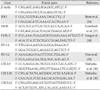
Total RNA was purified from hAd-MSCs using Trizol reagent (Invitrogen) according to the manufacturer's instructions. RNA samples were re-suspended in diethyl pyrocarbonate-treated water, quantified, and then stored at -80℃ until used. cDNA was synthesized from 1 µg of RNA using an reverse transcription polymerase chain reaction (RT-PCR) Kit (Intron, Seongnam, Korea). The PCR products after 35 cycles were subjected to electrophoresis on a 2% agarose gel. Bands were visualized under UV light and images were captured using a Multi-Image Light Cabinet (Alpha Innotech Corp., San Leandro, CA, USA). Primers used for RT-PCR are listed in Table 1.
Results
Establishment of hAd-MSCs
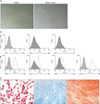
After initial expansion, cells were grown in EGM-2 or in a mixture of EGM-2 and Dulbecco's modified Eagle's medium (DMEM) at a ratio of 1:4. Cells cultured in EGM-2 proliferated faster than those cells in the mixed media, however, cells exhibited a similar spindle-shaped and fibroblast-like appearance, regardless of the culture media in which they were grown (Fig. 1A), and were adherent to the plastic culture surface, typical of human MSCs [31]. During passage 4-8, the cells were subjected to flow cytometric analysis for the identification of stem cell markers which revealed that cells were negative for hematopoietic markers such as CD34, CD45, and CD117 (Fig. 1B, upper panels), and positive for stem cell markers such as CD29, CD44, CD90, and CD105 (Fig. 1B, lower panels). There was no difference in markers expressed by the cells cultured in EGM-2 only and those cultured in the mixed medium (Electronic Supplementary Fig. 1).
Following the induction of cell differentiation using the commercially available cell-specific culture media, cell differentiation to adipocytes, chondrocytes, or osteoblasts was confirmed using Oil Red-O, Alcian blue, or Alizarin red S, respectively (Fig. 1C). Application of these staining methods to hAd-MSCs before culturing in differentiation medium showed no staining for these cell types (Electronic Supplementary Fig. 2). These results indicate that the cells obtained from adipose tissue were MSCs.
Inhibition of mouse T cell proliferation by hAd-MDC
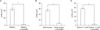
To confirm the well-known in vitro suppressive effects of MSCs on T cell proliferation, human PBMCs were stimulated with anti-human CD3 and CD28 antibodies and cultured in the presence or absence of hAd-MSCs at a ratio of 1:10 (hAd-MSC:PBMC). The presence of hAd-MSCs in the culture almost completely blocked the proliferation of human PBMCs (Fig. 2A). When mouse splenocytes (Fig. 2B) or isolated splenic T cells (Fig. 2C) were co-cultured with hAd-MSCs in the same way, similar results were obtained. Thus, Ad-MSCs of human origin suppressed the proliferation of mouse T cells, as well as of human PBMCs.
Activation of hAd-MSCs by murine T cell culture supernatant
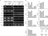
The aforementioned results also imply that hAd-MSCs were activated with mouse T cell supernatant, to exert suppressive effects on mouse T cell proliferation. When activated, MSCs up-regulate COX-2, iNOS, and/or IDO, through which they suppress T cell proliferation by secreting PGE2, NO, and/or IDO to the surrounding environment [6, 8, 9]. Additionally, they express PD-L1 to suppress T cells by contact [11]. Thus, we determined the expression of these molecules, as well as of chemokines, which are needed for recruitment of T cells. When the hAd-MSCs were treated with culture supernatant from PBMCs activated with various activators, they expressed all the molecules examined except iNOS (Fig. 3A, left panels). Meanwhile, when treated with mouse T cell supernatant, hAd-MSCs expressed immunomodulatory molecules differently depending on the mode of T cell activation, as is shown in the right panels of Fig. 3. Overall, the expression levels of the molecules, when it was expressed, were lower than those treated with the PBMC supernatant. Noticeably, COX-2 was expressed in all cases, but IDO was not expressed in any case. Densitometric values for each band were determined (Fig. 3B).
Inhibition of murine T cell proliferation by hAd-MSCs mainly via COX-2 expression
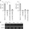
The above results indicate that the suppressive effect of hAd-MSCs upon mouse T cell proliferation may be principally due to PGE2, the product of COX-2. To verify this, NS-398, a COX-2 inhibitor, was added to the co-culture of mouse T cells and hAd-MSCs. With the addition of 0.5 mM NS-398, the suppression of T cell proliferation was almost completely recovered (Fig. 4A, left panel). Meanwhile, inhibitors of IDO (1-MT) and iNOS (L-NAME) did not affect the suppressive effect of hAd-MSCs on mouse T cell proliferation as expected (Fig. 4A, right panel).
The component in the mouse supernatant that stimulated hAd-MSCs was still not known. These cells are known to be critically activated by IFN-γ [32], a cytokine known not to be interchangeable between the human and mouse forms [25]. Thus, we assumed that TNF-α and/or Th2 cytokines, such as IL-4 and IL-5, in the murine supernatant would help activate these cells. To this end, hAd-MSCs were treated with mouse T cell culture supernatant or recombinant mouse cytokines, including TNF-α, IFN-γ, IL-4, and IL-5, and were harvested for RNA preparation. RT-PCR analysis revealed that murine T cell culture supernatant and TNF-α induced COX-2 expression in the hAd-MSCs (Fig. 4B).
Discussion
In the present study, we isolated MSCs from human adipose tissue. The cells were characterized by their adherence to the bottom of the culture dish exhibiting a fibroblast-like appearance; expression of surface markers and absence of hematopoietic markers; and their ability to differentiate into adipocytes, chondrocytes, and osteoblasts-all these characteristics attributed to cells defined as MSCs [31]. These cells, when treated with the culture supernatant of PBMCs, which are presumed to primarily be T cells, expressed COX-2 and IDO, through which the proliferation of PBMCs is suppressed. PD-L1 and PD-L2 were also expressed, which have also been shown to help the suppressive effect of the mesenchymal cells [11].
For the cultivation of hAd-MSCs, we used a mixture of EGM-2 and DMEM culture medium (1:5 ratio), in which the cells exhibited slower proliferation, but the same surface markers to those grown in only EGM-2 medium, and also exhibited the capacity to differentiate to adipocytes, chondrocytes, and osteoblasts. Considering that EGM-2 is expensive, and that mixture with DMEM did not deteriorate the stemness of MSCs, it would be beneficial to use the mixed media for the maintenance of MSC cultures.
When hAd-MSCs were stimulated with the PBMC supernatant, COX-2 and IDO were expressed, but not iNOS, consistent with previous reports [13, 26]. However, when these cells were stimulated with mouse supernatant, of the three genes designated as enzymes secreting soluble factors responsible for the immunosuppressive function of MSCs, only COX-2 was expressed (Figs. 3, 4). This finding implies that the manner in which mouse T cell supernatant stimulates hAd-MSCs differs from that of human T cell supernatant. To express IDO, human MSCs must be exposed to IFN-γ. When the PBMC culture media was applied to hAd-MSCs after incubation with an antibody to each cytokine, only that treated with the anti-IFN-γ antibody markedly decreased its capacity to induce the hAd-MSCs to express IDO (Electronic Supplementary Fig. 3A). Again, IFN-γR1-deficient human bone marrow (BM)-MSCs did not express IDO, even though they were co-cultured with PBMCs [32], indicating a critical role of IFN-γ signals for the expression of IDO. Our hAd-MSCs should be presented with murine IFN-γ when co-cultured with activated mouse T cells or when treated with the mouse supernatant; however, they failed to express IDO. This outcome is likely due to the fact that murine IFN-γ is not interchangeable with the human form [25, 33]. That is, even though the receptors of the two species show 53% homology, they hardly bind with IFN-γ from another species. Concordantly, hAd-MSCs expressed IDO when they were treated with human IFN-γ (Electronic Supplementary Fig. 3B), while they did not express IDO, when treated with murine IFN-γ (Fig. 4A).
Without IDO, hAd-MSCs still exerted in vitro suppressive effects upon the proliferation of mouse T cells. Undoubtedly, this effect is, at least in part, attributed to the expression of COX-2. Human BM-MSCs secrete PGE2, the product of COX-2, when cultured in the presence of IFN-γ or TNF-α [34], as was also shown in our results (Electronic Supplementary Fig. 3B). COX-2 was also induced by treatment with mouse supernatant and recombinant murine TNF-α (Fig. 4A). This was as expected, because murine TNF-α binds to both the human TNFαR1 and R2 TNF-α receptors [35]. It is also important to note that, although mouse TNF-α acts on human cells, a higher concentration than that needed for human TNF-α is required to obtain a similar effect [36]. This could explain the tiny COX-2 PCR bands obtained from the mouse supernatant or TNF-α treatment. Meanwhile, considering that the concentration of murine TNF-α that was used (1 ng/ml) is higher than that which would be found in the supernatant of cultured T cells, and that the intensity of the COX-2 band is weaker than that induced by the supernatant (Fig. 4B), it appears that TNF-α is not the sole factor to induce COX-2 in the hAd-MSCs. Other factors, currently unknown, could synergize the action of TNF-α.
In the murine environment, at least in vitro, COX-2 seems to be the main cause of the suppressive effect of hAd-MSCs on mouse T cell proliferation, because the COX-2 inhibitor NS-398 almost completely recovered mouse T cell proliferation (Fig. 4B, left panel), while the inhibitors of IDO (1-MT) or iNO (L-NAME) failed to recover the suppressed proliferation. Whether or not this is also the case in vivo is still unknown. Recent studies have suggested that factors other than IDO and COX-2 function as an immunomodulatory tool for MSCs. For example, increased production of adenosine via increased expression of CD39 on MSCs has reportedly contributed to the suppression of T cells [37]. In mouse MSCs, FAS ligand is expressed, and binds to FAS on T cells, thus leading to T cell death [38]. More importantly, MSCs express IL-10 and TGF-β [3, 4], cytokines that drive regulatory T cell production and reduce Th17 differentiation, both of which result in the diminution of T cell-mediated immune responses [39]. While IL-10 is not interchangeable between human and mouse species, TGF-β is [40]. Thus, it is highly plausible that TGF-β takes part in the immunomodulatory function of hAd-MSCs in experimental animal models. Even though we did not examine the secretion of TGF-β from hAd-MSC treated with mouse supernatant, experimental animal studies have reported both increased Treg and reduced Th17 production with human MSC administration for the treatment of disease models [15, 16, 22, 41].
In conclusion, hAd-MSCs undergo different modes of activation, when they were treated with mouse T cell culture supernatant, compared to when they were stimulated with human PBMC supernatant. However, they still exerted an anti-proliferative effect on mouse T cells in vitro, primarily through COX-2 expression. These results have not previously been reported, and should be considered when interpreting the results of experiments evaluating the effects of hAd-MSCs in experimental animal models. More sophisticated studies are needed to analyze the exact mechanisms of immunomodulation by hAd-MSCs in experimental disease models.
 XML Download