

 PDF
PDF ePub
ePub Citation
 Print
Print


Introduction
The brachial plexus is a complex network of nerves originating in the neck that supplies the upper limb. It courses through the axilla and is formed by the anterior primary rami of spinal nerves C5, C6, C7, C8, and T1. The C5 and C6 roots unite to form the upper trunk, the C7 root continues as the middle trunk, and the C8 and T1 roots join to form the lower trunk. Each trunk divides into ventral and dorsal divisions. The ventral divisions of the upper and middle trunks join to form the lateral cord, and the ventral division of the lower trunk forms the medial cord. The dorsal divisions of all three trunks join to form the posterior cord [1, 2]. Upper limb buds develop as outgrowths from the ventrolateral body wall opposite the lower five cervical and upper two thoracic segments. As the ventral rami of these segments rearrange themselves to form the brachial plexus, anatomical variations may occur [3]. Understanding these variations is important for clinicians, as the plexus may be vulnerable to injury during radical neck dissection, axillary lymph node clearance for malignancies, shoulder arthroscopy, and infraclavicular nerve blocks [4]. In this report, an extremely rare case of bilateral occurrence of the brachial plexus united into a single cord is described and its potential implications are discussed.
Case Report
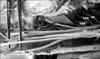
The anatomical variations in the present case were observed on routine dissection of an elderly female cadaver, approximately 65 years of age, of South Indian origin. The brachial plexus on the right and left sides of the cadaver was dissected. The supraclavicular, pectoral region, axilla, and arm were dissected. The roots, trunk, cords, and branches of the brachial plexus were identified, and their relationships with the axillary artery were noted.
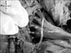
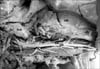
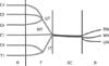
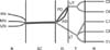
On the left side, C4, C5, and C6 roots combined to form the upper trunk, the C7 root formed the middle trunk, and C8 and T1 roots combined to form the lower trunk. All three trunks almost immediately fused to form a single cord (Fig. 1), which was lateral to the subclavian artery in the supraclavicular region. All the branches of the brachial plexus that typically arise distal to the roots branched off from this pre-fixed common cord (Fig. 2). The long thoracic nerve arose from C5, C6, and C7 nerve roots, as in normal anatomy. All the branches that arose from the common cord were either supra- or retroclavicular in origin. This was noted during dissection of the axilla, where only the branches of the brachial plexus, but not the cords, were noted. Fig. 3 is a schematic diagram showing the variations that were observed on the left side.
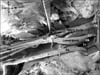
On the right side, C5 and C6 roots formed the upper trunk, which divided into anterior and posterior divisions (Fig. 4). C7, C8, and T1 roots combined to form the lower trunk. The middle trunk was absent. In the supraclavicular region, the posterior division gave rise to three branches, the suprascapular nerve and the upper and lower subscapular nerves. The anterior division then joined the lower trunk. This was rejoined by the posterior division of the upper trunk after it gave rise to the aforementioned branches to form the single cord (Fig. 5). All the remaining branches of the brachial plexus arose from this single cord (Fig. 5). Similar to the left side, this cord was not seen in the axilla, where only the branches were observed. A schematic representation of the variations noted on the right side is shown in Fig. 6.
On both sides, the fused cord lay superior to the subclavian artery in the posterior triangle of the neck, anterolateral to this artery while descending deep to the clavicle, and finally lateral to the axillary artery in the axilla. No other anomalies were noted in the cadaver.
Discussion
An extensive literature search using PubMed, Google Scholar, and a general Google search revealed only nine previously reported cases of the brachial plexus united into a single cord (Table 1). To the best of the authors' knowledge, bilateral single cords have not been described in any of these cases. In another recent study from India, four cases (4.5%) of a single cord were reported among 45 cadavers (90 brachial plexuses), two on the right side and two on the left [5]. The findings of this study are particularly unusual as no other large study of the brachial plexus has described the formation of a single common cord [6, 7]. All other publications on single common cords are case reports [8-10]. In two cases, the upper trunk (fused C5 and C6 roots) united with C7, C8, and T1 roots to form the single cord. In one instance, the formation of the upper, middle, and lower trunks followed the usual pattern. The upper trunk divided into anterior and posterior divisions, and the middle and lower trunks, without dividing, directly fused with the two divisions of the upper trunk to form a single cord. In the last case, the upper, middle, and lower trunks were formed as usual, after which they directly fused to form a single cord, as in the present case on the left side [5]. Similar findings, in which the contribution of the C4 root was evident, were reported in a case from India published over 50 years ago [10].
Another case report from India described the formation of an infraclavicular common cord of the brachial plexus formed by fusion of divisions of all the three trunks [8]. The branches that usually arise from the cords also arose from the common cord [8]. Only three case reports of a single cord have been reported in the Western literature [9]. All these case reports were published in the first half of the 20th century (Table 1). It is notable that cases of single cords have been more commonly reported from India, in males, and on the left side. This warrants further study with larger sample sizes.
Variations in the anatomy of the brachial plexus may be attributable to factors that influence the formation of limb muscles, peripheral nerves, or associated vessels during the embryonic period. Embryologically, the brachial plexus appears as a single radicular cone of axons of spinal nerves, growing distally to reach the muscles and skin of the upper limb, which later divide to form the ventral and dorsal divisions [11]. Sannes et al. [12] suggested that guidance of the developing axons is regulated by expression of chemoattractants and chemorepulsants in a highly coordinated site-specific fashion. Several signaling molecules and transcription factors have been identified, which induce the differentiation of the dorsal and ventral motor horn cells. Misexpression of any of these signaling molecules can lead to abnormalities in the formation and distribution of particular nerve fibers [13]. Variation in patterns of formation and branching of the brachial plexus from species to species is supported by phylogenic evidence. No trunk formation is seen in amphibians, reptiles, or dogs, whereas in gorillas, two trunks are formed, with C4, C5, and C6 roots forming the first trunk and C7, C8, and T1 roots forming the second trunk [14].
It was noted with interest that in all reported cases of a single cord, the subclavian artery was located inferior to the cord. More distally, the axillary artery was medial to the cord. The subclavian artery usually arises from the seventh cervical intersegmental artery, which intervenes between the middle and lower trunk of the brachial plexus. Distally, it continues as the axillary artery and is surrounded by the cords of the brachial plexus. In the case of a single common cord, the subclavian artery possibly arises from a more distal intersegmental artery rather than the usual seventh cervical intersegmental artery. This would allow for the more proximal fusion of the constituents of the brachial plexus, and accordingly, an artery would no longer intervene between the middle and lower trunk [3, 15].
The clinical implications of a single common cord are not certain because of its rarity. However, it is likely that any supraclavicular brachial plexus injuries in such individuals would have serious clinical manifestations as all the major branches of the brachial plexus arise from this common cord. Additionally, the single common cord variant would pose difficulties in the event that individual cords need to be selectively blocked [5]. In addition, structures might be misidentified during surgeries of the cervical spine, especially in the case of nerve sheath tumors such as schwannomas [16]. The altered relationship of the subclavian artery to the common cord is likely to reduce the probability of the brachial plexus being affected as a result of pathologies or interventional procedures of the subclavian vessels.

 XML Download