

 PDF
PDF ePub
ePub Citation
 Print
Print


Introduction
The liver and the gallbladder are the main parts of the hepatobiliary system. They are supplied by the branches of the celiac trunk, one of the 3 ventral branches of the abdominal aorta. The celiac trunk terminates by dividing into the left gastric, splenic, and common hepatic arteries. The left gastric artery supplies the stomach; the splenic artery supplies the stomach, spleen, and pancreas; and the common hepatic artery supplies the stomach, duodenum, pancreas, and the hepatobiliary system. The common hepatic artery normally terminates by dividing into the gastroduodenal and proper hepatic arteries. The gastroduodenal artery divides into the superior pancreaticoduodenal and right gastroepiploic arteries, while the proper hepatic artery provides the right gastric artery and then divides into the right and left branches, which supply the right and left liver lobes. The right hepatic artery provides a cystic branch, which passes through Calot's triangle and supplies the gallbladder. Many variations of the branching pattern of the celiac trunk have been previously described. In the very rare cases in which the celiac trunk is completely absent, all its branches arise from the abdominal aorta [1]. The hepatogastric trunk, splenic artery or hepatosplenic trunk, and the left gastric artery have been reported to directly originate from the abdominal aorta [2]. Other variations of the celiac trunk, such as presence of a celiacomesenteric trunk [3] and celiacomesenteric-phrenic trunk [4] have also been described. A thorough knowledge of possible variations of branching, course and distribution of the vessels supplying the liver and gallbladder is essential during laparoscopic surgeries of these organs, and is also important for radiological procedures and therapeutic embolization. We found several rare variations of the hepatic and cystic vessels that could be significant for various clinical disciplines. The aim of this case report is to alert clinicians to the existence of vascular variations of the liver and the gallbladder that could result in fatality during procedures.
Case Report
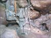
We noted variations of the hepatic and cystic arteries in a male cadaver (approximate age, 70 years) during routine dissection classes for undergraduate medical students. The celiac trunk trifurcated into the splenic, left gastric, and common hepatic arteries. The common hepatic artery trifurcated into the right hepatic, left hepatic, and gastroduodenal arteries, 2.5 cm above the first part of the duodenum. The right gastric artery arose from the left hepatic artery 5 cm above the level of the pylorus. The right hepatic artery divided into a left and right branch after a course of 2.5 cm. The left branch entered the right hepatic lobe through the porta hepatis. The right branch passed to the right behind the common hepatic duct (between common hepatic duct and the portal vein) to reach Calot's triangle, where it provided 2 cystic branches to the gallbladder and entered the right lobe of the liver through the porta hepatis. The common hepatic duct was 6 cm in length and joined the cystic duct immediately above the level of the first part of the duodenum (Fig. 1).
Discussion
Vascular variations of the liver and gallbladder are quite common. In the present case, we encountered a unique variation in the celiac trunk branches. The common hepatic artery trifurcated into the gastroduodenal, right hepatic, and left hepatic arteries. The right hepatic artery divided into right and left branches, of which the right had a variant course and branches. Although many reports have been published on the variant branches of the hepatic artery, the variation we report here has not been previously described. Abdullah et al. [5] extensively studied the variations of the hepatic arteries in liver transplant cases. In their study of 932 patients, variations of the hepatic artery were detected in 31.9%. Of these, 48 cases had common hepatic artery anomalies, and 236 cases had left or right hepatic artery anomalies. These investigators also noted 13 rare variations, including 1 case of right hepatic artery stemming from the inferior mesenteric artery and 1 case of a normal common hepatic artery passing behind the portal vein. Uva et al. [6] reported a case of common hepatic artery arising from the left gastric artery. Song et al. [7] studied the common hepatic artery in 5002 patients and found variations in only 3.71% cases. In this series, the common hepatic artery arose from the left gastric artery in 0.16% cases and passed into the liver through the fissure for ligamentum venosum. It arose from the superior mesenteric artery in 3% and from the abdominal aorta in 0.40% of their patients. Okada et al. [8] also found that the common hepatic artery originated from the left gastric artery in 3% cases. Further, Mishall and Rajgopal [9] have reported a case of the right hepatic artery forming a caterpillar hump.
The cystic artery is normally a branch of the right hepatic artery that passes posterior to the common hepatic duct to reach the neck of the gallbladder. It may originate from the right hepatic (63±9%), hepatic trunk (26±9%), left hepatic (5±5%), gastroduodenal (2±6%), superior pancreaticoduodenal (0±3%), right gastric (0±1%), celiac trunk (0±3%), or superior mesenteric (0±8%) arteries [10]. Mlakar et al. [11] studied the blood supply of the gallbladder in 81 subjects, and found that the gallbladder was supplied by 1 cystic artery in 86% and by 2 arteries in 14% cases. When a single artery was present, it originated from the right hepatic artery in 53% livers. In another study [12], the cystic artery was most often a single vessel (97.06%) and rarely a double vessel (2.94%). It arose most often from the right proper hepatic artery (82.34%), rarely from its trunk (8.82%) or its left branch (5.88%) and most rarely from the gastroduodenal artery (2.94%). The incidence of double cystic artery is 2-15%. In such cases, ligation of both cystic arteries may become necessary during cholecystectomy for treatment of gallbladder stones, inflammation, edema, adhesions, and fibrosis [11-13].
In the present case, the celiac trunk trifurcated into the gastroduodenal, left hepatic, and right hepatic arteries. The right gastric artery arose from the left hepatic artery. A recent report describes variation of the hepatic vessels in the setting of multiple vascular and ductal anomalies of the liver [14]. Knowledge of this variation may be important for surgeons performing laparoscopic cholecystectomy, therapeutic embolization of the hepatic arteries or other radiological interventions in this area [15]. The bifurcation of the right hepatic artery, its course in Calot's triangle and the presence of double cystic arteries are unique features of this case. Calot's triangle was also large owing to the low union of the common hepatic and cystic ducts. Ligation of the right branch of the right hepatic artery in Calot's triangle during cholecystectomy could cause hypoxia in the part of the liver it supplies. The large size of Calot's triangle in this case would increase the chance of ligating this artery, without its distribution to the liver being noticeable. Even if the right branch of the right hepatic artery is identified and its ligation avoided, ligation of only 1 of 2 cystic arteries can result in significant hemorrhage from the other artery.
A thorough knowledge of the branching pattern of the hepatic and cystic arteries is surgically important. Insufficient recognition of anatomical variation of these vessels may create a dangerous situation, especially during laparoscopic cholecystectomy. When the variations are multiple, as in the case reported here, the chances of vascular damage are very high. It is therefore advisable to conduct a radiological or tomographic evaluation for possible vascular variations when planning any surgery of the liver or gallbladder.
 XML Download