

 PDF
PDF ePub
ePub Citation
 Print
Print


Introduction
Evidence of trauma is one of the most frequently observed medical signs in human skeletons from archaeological sites [1-4]. Osteological studies on traumatic injuries have recently evolved from simple case reports to in-depth, population-level investigations [5, 6]. Some studies have extended this line of inquiry further, investigating trauma patterns in association with socioeconomic conditions. In the process, medical knowledge of long-bone fractures in history has become much more comprehensive [1, 3, 7, 8]. Categories of traumatic injuries in historical skeleton samples include fracture, dislocation, and post-traumatic deformity. [2, 4]. Long bone fractures have been studied over the past several decades. The earliest trauma samples discovered in archaeological sites are Homo erectus bones taken from a Choukoutien cave in China [4, 9], as well as Homo sapiens specimens dating to the Paleolithic through Neolithic periods [9, 10].
The historical literature suggests that fracture hazards are usually determined by cultural, environmental, and socioeconomic conditions prevailing in a given human society. The prevalence and distribution of fractures change with the culture and subsistence base of each human society [2, 4, 7, 10-15]. Securing data from as wide a geographic and temporal range as possible is desirable to expand the paleopathological knowledge base on fractures. However, research on the variances in fracture prevalence relative to temporal and geographic conditions is inadequate. In East Asian countries, where industrialization was delayed until recently, no studies have been conducted on fractures in premodern societies yet.
Over the past several years, we attempted to build a human skeletal sample collection consisting of specimens obtained from 16th-18th century Joseon tombs in Korea. Our investigations of the collection have revealed important information concerning the health and disease status of pre-modern Joseon people [16-21]. We undertook osteological examinations of skeletons from the same collection to determine the prevalence and location of long bone fractures in premodern Korean specimens. The purpose of our study was to contribute to a better understanding of the fracture patterns prevailing in a premodern East Asian population.
Materials and Methods
We examined the long bones of Joseon Dynasty skeletons, maintained at Seoul National University College of Medicine, Korea, to identify osteological evidence of fractures. Archaeological evidence and carbon dating results confirmed that every skeleton in this study belonged to 16th-18th century Korean people. Although the cases examined in this series were collected from different cemeteries of Joseon period, the tombs that contained these skeletons were of the same type, Hoegwakmyo. This type of Joseon tomb was surrounded by a fortified barrier made of a lime-soil mixture that successfully reduced grave intrusion and robbery.
The long bones examined (n=96) included the humerus, ulna, radius, femur, tibia, fibula, and clavicle. Since 61 bone elements (4.5%) were lost or severely damaged, the total number of elements examined was 1,283. We determined the sex of the individuals by the shape of the sciatic notch or mastoid process. We estimated the age at death following the method of Lovejoy et al. [21]. Briefly, individuals were classified into adolescent (12-20 years), young adult (20-35 years), middle aged (35-50 years), and old (>50 years) groups using osteological indicators such as the degrees of transverse organization, granularity, apical activity, retroauricular activity, and articular surface porosity.
Genuine fractures were differentiated from post-mortem damage after interment by suggestive clues such as the surface coloration in the fracture site distinct from other parts [2, 4, 22]. Affected and unaffected sides of long bones were always compared in the same individual because rotation or abnormal deviation of the bone could not be easily identified in the cases that exhibited other pathologies such as infections [4]. We also determined the type of long bone fractures. Direct trauma causes penetrating, comminuted, transverse, and crush type fractures, while indirect trauma causes spiral, oblique, torus, impacted, burst, and avulsion fractures [2]. Of these, transverse and oblique fractures are especially significant, because transverse fractures are known to result from intentional violence, whereas oblique fractures are mainly sustained in accidents [4]. The position of fracture in the shaft of the long bone, i.e., proximal, middle or distal third, was recorded following the method of Grauer and Roberts [7]. Rotational or linear deformity, healed fracture alignment, bone shortening, presence of osteomyelitis, and callus formation were also examined and described.
In some disputed cases, the pattern of fracture was confirmed by radiographic analysis. Following gross inspection, simple radiographs of the fracture were obtained in standard anteroposterior and lateral views. Radiographs in the mediolateral or tangential views were also obtained if necessary. Percentage of fracture was calculated for each element and individual. Multiple fractures were defined by presence of ≥2 fractures, and were calculated for each individual.
To presume the good reasons for arm fractures of Joseon people, we also searched for information in The Diaries of the Royal Secretariat (Seungjeongwon Ilgi), the historical literature of the Joseon Dynasty [23].
Results
The study population included 96 individuals: 56 men (58.3%) and 40 women (41.7%). Of these, 5 individuals (cases nos. 33, 41, 65, 98, and 100) showed fractures (5.2%). The number of bone elements fractured was 7 (0.55%) (Table 1). All of them exhibited signs of healed fractures, but none had evidence of post-mortem changes after interment. Four fractures were found in the upper extremity bone elements, while 3 fractures were identified in the lower extremity elements (Table 1). Fractures were identified in middle-aged (cases nos. 33, 98, and 100) and old (cases nos. 41 and 65) individuals. Four fractures were identified in men (cases nos. 41, 65, 98, and 100) and only 1 in women (case no. 33). Oblique fractures were observed in 6 elements (cases nos. 33, 65, 41, and 100), while a transverse fracture was found only in the left ulna of case 98 (Table 1).
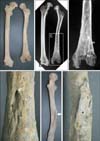
In case no. 33 (middle-aged woman), an oblique fracture was identified in the middle of the left femoral shaft. Marked deformity, axial deviation, and rotation were associated with shortening of the bone length after fracture (Fig. 1A). The marked changes observed on gross examination of the femur suggested that the healing process was atypical. Radiography showed bony irregularities in the medullary cavity (Fig. 1B, C). We also found an opening in the posterior part of the left femur, possibly representing a cloaca penetrating into the marrow space (Fig. 1D). A bony fragment, possibly healed in a malaligned position, was identified on its anterior surface (Fig. 1E, F). All these signs suggest that the individual might have had osteomyelitis as a complication after femur fracture.
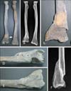
Case no. 41 (old man) showed a Colles' fracture in the distal right radius; fracture type was oblique. We observed radial shortening on gross examination (Fig. 2A, B) with dorsal and proximal displacement of the distal fracture fragment (Fig. 2A, D). The distal radius also showed radial angulation with cortical disruption and a sharp edge on the volar side, suggesting that healing occurred without proper reduction (Fig. 2C, E). Callus formation, dorsal tilt, and shortening of the bone were prominent radiographic features in the right radius (Fig. 2F). Case no. 41 probably experienced significantly limited range of motion and loss of function because of the severe angulation and bone length shortening.
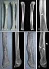
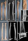
Case no. 65 (old man) had fractures in the distal left fibula and tibia, with bony thickening in the distal tibia (Fig. 3A). An oblique fracture line was observed on radiographs of the tibia (Fig. 3B, C). The distal left fibula was also thickened and rotated, and radiographic examination revealed a radiolucent fracture line and cortical thickening (Fig. 3D, G). In case no. 98 (middle-aged man), a transverse fracture was noted in the mid-shaft of the left ulna (Fig. 4A, C), most likely caused by direct intentional force. The lower left ulnar shaft was widened without shortening of the length (Fig. 4A, B). Radiographs showed the radiolucent fracture line and callus formation (Fig. 4B, E). No evidence of malalignment or rotation of the ulnar axis was observed.
Case no. 100 was a middle-aged man with oblique Colles' fractures in both radii. The left radius was shortened (Fig. 4F). Radiographs showed prominent osteosclerotic change in the right radial metaphysis, suggesting recent fracture (Fig. 4G, H). Dorsal angulation was also confirmed by the presence of volar tilt on the lateral view, measured as the angle created by articular surface of distal radius and a line drawn perpendicular to the long axis of the radius (Electronic Supplementary Fig. 1). The distal angulation between the long axis of the radial bone and the distal end of the radius was more prominent in the left radius. Although the fractured area was covered by remodeled bone, significant proximal displacement was not evident (Electronic Supplementary Fig. 1C, D).
Discussion
In this study, among the many factors possibly influencing fracture frequency, we first considered age and sex. All of our long bone fracture samples were from middle-aged or old individuals. This is understandable, since the long bones from older individuals are likely to be more fragile and prone to injury by even low-energy impacts. Senile osteoporosis is known to significantly increase the vulnerability of bones to trauma [4]. We also observed that long bone fractures were much more common in men than in women. As noted in studies on other human populations [24, 25], this male preponderance might be associated with typical sex-related differences in physical activity. Joseon men typically performed more strenuous work than women, and so would have sustained a higher frequency of fractures.
We also considered the social and economic status of the individuals in our interpretation of the current results. Most skeletons examined in this study were collected from Hoegwakmyo, a unique burial system of the Joseon Dynasty. The people of Joseon society regarded Hoegwakmyo as an ideal tomb because this manner of constructing tombs was highly recommended by Chu Hsi, the grandmaster of Confucianism [26]. Nevertheless, the tomb was only adopted by the rich Joseon elites because its construction was too expensive for lower-class individuals. Therefore, when archaeologists in Korea found Hoegwakmyo tombs in archaeological fields, they assumed that the human remains in these tombs must be from the upper class of Joseon society [26, 27]. Most of the individuals represented in the present study are much more likely to have been landowners than farmers [26].
The fracture type and location, also known to vary by population [14, 15], were also considered in this study. Although a number of investigators have reported a high frequency of clavicular involvement in fractures [13, 28, 29], we could not identify any clavicle fractures in our Joseon skeletal series (Table 1). The pattern of forearm fracture in the present study was unique. In most previous studies, a very high percentage of fractures occurred in the forearm bones, with the ulna broken more often than the radius [10, 29, 30]. One such example is the Monteggia fracture, a fracture of the proximal third of ulna with dislocation of the radius head. Paleopathologists consider the finding of a large number of Monteggia fractures in a skeletal series important because they may have been incurred while fending off a blunt attack, implying that the people lived in an environment where violence was rampant [1, 14, 30]. Although forearm fractures were commonly observed in the present study, each 1 involved the radius, with the exception of 1 element (case no. 98, possibly a Monteggia fracture). We discovered 3 Colles' fractures of different severity in 2 individuals (cases nos. 41 and 100) (Table 1).
The Colles' fracture is generally associated with indirect, accidental falls on the hand (e.g., a short fall caused by a shift in body weight), resulting in angulation, proximal displacement of the distal fragment, and bone length shortening [28, 31, 32]. Similarly, accidents can be considered the cause of the Colles' fractures in this study. The high-class, elite Joseon individuals buried in Hoegwakmyo would naturally have fewer incidents of hard-labor-related accidents that could have caused bone breakage. Some, although not all, Colles' fractures in this study might have been caused by accidents characteristic only of the upper classes in Joseon society. Our search for contemporary articles on fractures in historical documents of Joseon society (Seungjeongwon Ilgi) interestingly showed that many arm injuries of the Joseon elite were caused by accidents such as a fall from a horse (Electronic Supplementary Table 1).
The fracture types also indicate a rarity of injuries possibly induced by intentional violence in our series. Most of the fractured elements were of the oblique type (6/7, 85.7%), suggesting indirect, accidental forces acting from a distance [2, 7]. Transverse fracture, typically associated with intentional violence [4, 33], was identified in only 1 individual (case no. 98). This supports the conclusion that the Joseon individuals examined in this study might not have been commonly subjected to blunt trauma in a violent setting. We can assume that the Joseon elite spent tranquil lives.
Post-fracture complications include infection, tissue necrosis, abnormal or non-union, and bone deformity. Deformations can develop or worsen because individuals living with fractures compensate for their injuries [4]. We observed a variety of fracture-induced complications in this study, for example malalignment (cases nos. 41, 98, and 100) caused by incomplete reduction of the injury. In case no. 33, post-fracture infection appeared to have extended to the medullary space, and this is particularly interesting because although osteomyelitis was a common disorder in centuries prior to the use of antibiotics, it has rarely been traced in archaeological samples [4, 11, 30]. Because of the difficulty in diagnosing osteomyelitis by bone examination alone, researchers worldwide hesitate to definitively declare lesions to be osteomyelitis without very clear, incontrovertible supporting evidence [4]. Our case no. 33 may be 1 such rare example, in which we confirmed the most typical osteological signs of osteomyelitis, including bony proliferation, cloaca opening, and bone marrow involvement.
The unique points of our study were not limited to fracture type and location. Skeletal series in most countries have revealed fracture rates of 15.1-32.9% (person fracture rates) and 2.4-11.7% (total % of fractured elements). However, the long bone fractures rates in our Joseon sample were only 5.2% (person fracture rate) and 0.55% (total % of fractured elements) (Electronic Supplementary Table 2). The fact that the samples in our skeletal series were collected only from the upper and ruling classes of the Kingdom and not from all strata of Joseon society, may explain the relatively low fracture prevalence observed in this study. The low fracture rate might well have reflected the less strenuous physical labor of the Joseon individuals examined, since performance of heavy physical labor is closely associated with fracture frequency [29]. However, the comparison of disease signs in different skeletal series must be approached discreetly if the research methods used have not been sufficiently standardized [34]. Definitive conclusions cannot be derived while the technical limitations inherent in non-standardized studies on skeletal series persist under different conditions.
Despite its shortcomings, the present study is of interest to scientists in related medical fields. The skeletal series of East Asian societies might manifest unique fracture patterns because of the distinctive lifestyle that they have maintained for many millennia. However, few simple descriptive studies have been conducted on skeletons collected from archaeological fields in Korea or other East Asian countries until now. Our study involves long bone fracture cases evidently representing a 16th-18th century Joseon skeletal series. We have presented the first description of some unique points of fractures observed in these 16th-18th century Joseon skeletons. Further research focusing on different social strata and populations is nevertheless required to gain a clearer understanding of trauma in historic Asian populations.

 XML Download