

 PDF
PDF ePub
ePub Citation
 Print
Print


Introduction
Morphine is an opiate narcotic analgesic commonly prescribed to treat moderate to severe pain. As a derivative of opium, morphine can lead to psychological and physical dependence and addiction. There are many surveys that focus on the functional and biochemical changes of the nervous system after the consumption of morphine, heroin, alcohol, marijuana, and cocaine that might be lead to addiction [1-6]. However, the structural and especially the morphometrical changes have received less attention, and the results of these studies are controversial [6-11]. Zhang et al. [12] explained that the protective and destructive roles of morphine in the neuronal system (neurons and glial) are unclear. There is evidence suggesting that morphine induces apoptotic cell death in neuronal and glial cells, whereas controversial studies support a neuroprotective role for morphine [12]. Tamura et al. [13] showed the neuroprotective effects of morphine on low-temperature-induced cell death. They reported that the neuroprotective effects of morphine were antagonized by naloxone, which is a non-selective opioid receptor antagonist [13]. Meanwhile, Hu et al. [14] reported that morphine induces apoptosis of human neurons and microglia and that naloxone blocked morphine-induced apoptosis.
In addition, the reports on the effects of morphine withdrawal on the structure of the nervous system are controversial and have not received enough attention [15, 16]. The reports on the changes in the glial cells are mainly focused on the qualitative, biochemical, and physiological alterations, but not the quantitative alteration, after drug use and abstinence [17-19].
The present study has focused on the neurons and oligodendrocytes of the medial prefrontal cortex (MPFC). The MPFC plays a major role in organizing and motivating addiction-related behavior [20, 21]. Alterations in the MPFC function may actually be associated with a predisposition to addiction [20, 21]. The present study aimed to investigate the effects of chronic morphine treatment (30 days) with or without abstinence (30 days) on the number of the neurons and oligodendrocytes in the MPFC structures. Modern stereological studies rely on the geometrical and statistical analyses and provide accurate and comparable results in quantitative research including neuropathology [22].
Materials and Methods
Animals
Twenty-four male adult Sprague Dawley rats, each weighing 250-300 g at the beginning of the experiment were randomly selected from the Laboratory Animal Center of Shiraz University of Medical Sciences (Shiraz, Iran). They were divided into four groups, each including six animals (n=6) that were acceptable for stereological survey [23]. The animals were housed under controlled humidity and temperature conditions in a 12-hour light/dark cycle with free access to food and water. Experimental protocols were approved by the Animal Ethics Committee of the Shiraz University of Medical Sciences by agreement No. 89-5431 (Shiraz, Iran).
Morphine treatment and abstinence
In this experiment, each of the animals were assigned to one of the four groups: 1) the control animals were treated with saline for 30 days (SAL); 2) the second group was chronically treated with morphine for 30 days (MOR); 3) the other control group was treated with saline for 30 days followed by another 30 days of abstinence injection (SAL+ABS); and 4) the last group was chronically treated with morphine for 30 days followed by 30 days of abstinence injections (MOR+ABS). Morphine sulfate (purchased from Temad Factory, Tehran, Iran) was dissolved in sterile saline and administrated subcutaneously at a dose of 1 ml/kg once daily for 30 days with an escalating dose of morphine. Briefly, the initial dose administered was 5 mg/kg and was increased by 5 mg/kg every five days to a maximum dose of 20 mg/kg, and continued until day 30 of the treatment [24]. The control rats received an equal volume of saline.
Tissue preparation
The animals were anesthetized with ketamine (100 mg/kg, i.p.) and xylazine (10 mg/kg, i.p.). Then, the rats were perfused transcardially. Briefly, the rat brains were fixed by cardiac perfusion with 0.9% saline followed by 200-300 ml of 4% paraformaldehyde in 0.1 M phosphate buffer, pH 7.4. Then, the brains were rapidly removed from the skulls, placed in buffered formaldehyde, and embedded in a paraffin block. A complete series of coronal sections were obtained, according to Paxinos and Watson [25]. The thickness of the sections was 25 µm, and they were stained with cresyl violet [26, 27].
Estimation of the volume of the MPFC and its subdivisions
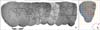
The brain was executively sectioned. Nine to eleven sections were sampled in each rat in a systematic, random manner, thus with a random start and equidistant. The volume of the right MPFC and its subdivisions (anterior cingulate, prelimbic, and infralimbic areas) were estimated using the Cavalieri method (Fig. 1) [22, 28-30]. The boundary of the MPFC was considered to be between the first appearance of the forceps minor of the corpus callosum (Bregma 4.20 mm) and its decussation (Bregma 2.28 mm) [20]. The border of each region was determined at a final magnification of ×16. A grid of points was superimposed on the sampled sections (Fig. 1). This test system of points was overlaid on the image of the tissue using stereology software designed at our research center (Morphometry & Stereology Research Center, Shiraz University of Medical Sciences, Shiraz, Iran). The estimation was made using the following formulas:
in which "V(MPFC)" is the volume of the cortex, "t" is the distance between the sampled sections and the section thickness, "ΣPi" is the total number of points hitting the sections of MPFC and its subregions, and "a(p)" is the area per point and is calculated by multiplying "ΔX" by "ΔY" (which, in this case, was 0.2 mm2). The right upper corner of each cross was considered a point (Fig. 1).
Estimation of the numerical density and the total number of neurons and oligodendrocytes
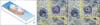
Microscopic survey was done using a videomicroscopy system made up of a microscope (E-200, Nikon, Tokyo, Japan), a video camera, a high numerical aperture lens (×60, N.A.=1.4), and a computer and a monitor. Ten to twelve microscopic fields were studied in each rat. The microscopic fields were selected in a systematic random manner, thus with a random start and then at an equal traveling distance. Briefly, a microscopic field was selected from a corner of the slide out of the MPFC. The slide was moved at equal intervals along the X- and Y-axes using a stage micrometer. This method was continued until the whole MPFC was quantified. The numerical density of the neurons and the oligodendrocytes was estimated according to the disector ("two sections") principle within the MPFC [22, 28-30]. The oligodendrocytes were identified using staining, according to Chareyron et al. [27], who explained that oligodendrocytes are smaller than astrocytes and contain round, darkly staining nuclei that are densely packed with chromatin. To estimate the total number of cells, an oil immersion lens at a magnification of ×3,500 was used. A brief description appears under Fig. 2. The numerical density of the cells was estimated using the optical disector and the following formula: in which "a(frame)" was the area of the counting frame (in this case, 736 µm2), "h" is the height of the optical disector (in this case, 10 µm), "ΣQ-" is the number of the cells counted in all disectors, "ΣP" is the total number of counted frames, "T" is the microtome setting to cut the block (in this case, 25 µm), and "t" is the real thickness of the section measured in three random areas of each microscopic slide using the microcator (MT 12, Heidenhain, Germany). In each field, the first 5 µm of the section thickness was ignored to avoid unbiased counting. This has been called a "guard zone" (Fig. 2). Cell counting was done in the next 10 µm of section thickness, which is known as "the height of the disector" or "h" (Fig. 2). The cells that were completely or partly inside the counting frame or touching the upper and right borders were counted, whereas the cells that hit the lower and left borders were not counted. In other words, just the cells whose nucleoli did not appear in the beginning of the disector height and appeared at the following optical scan were counted [22, 28-30]. The cells whose nucleoli were completely or partly inside the counting frame or touching the upper and right lines were counted "ΣQ-" (Fig. 2). "Reference trap" indicates that relying on the density (and not the total amount of the parameter) might lead to false conclusions; therefore, the numerical densities were multiplied by the MPFC volume [22, 28-30] .

Results
Volume of the MPFC
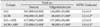
The volume of the MPFC and its subdivisions was decreased by approximately 15% in the MOR group compared to that in the SAL group (P<0.05). In addition, the volume decreased by approximately 24% in the MOR+ABS group compared to that in the SAL+ABS group (P<0.05) (Table 1).
Number of cells
The total number of neurons significantly decreased by approximately 44% in the MOR group compared with that in the SAL group (P<0.01). It significantly decreased by approximately 35% in the MOR+ABS group compared with that in the SAL+ABS group (P<0.01) (Table 1).
The total number of oligodendrocytes significantly decreased by approximately 41% in the MOR group compared with that in the SAL group (P<0.05). It decreased by approximately 37% in the MOR+ABS group compared with that in the SAL+ABS group (P<0.05) (Table 1).
No significant difference was seen in the number of the cells in the MOR and MOR+ABS groups.
Discussion
This study used stereological methods with the aim of evaluating the effects of chronic morphine consumption and abstinence on the neurons and oligodendrocytes in the MPFC of rats. A review of the literature showed that the results of the destructive or protective effects of morphine are controversial. Hodgson et al. [31] and Mao et al. [32] reported that intrathecal consumption of morphine might induce neurotoxicity in animals. In a study by Atici et al. [33], chronic administration of morphine was reported to cause apoptosis of neurons in the parietal, frontal, temporal, occipital, entorhinal, pyriform, and hippocampal regions of the brains of rats. In addition, an in vitro study showed that morphine causes increased neurotoxicity in human neuronal cultures with apolipoprotein E [34]. Some data indicate that morphine, through the activation of opioid receptors, can promote abnormal programmed cell death by increasing the expression of pro-apoptotic protein and suppressing the expression of anti-apoptotic protein [35]. Meanwhile, there are some reports of the protective effects of morphine. For example, the protective effects of morphine in hypoxia and ischemia have been reviewed by Peart et al. [36]. In addition, Rambhia et al. [37] suggested that morphine induces a neuroprotective effect mediated by nitric oxide in a human neuroblastoma cell line against intracellular oxidative stress and neuroinflammation. In addition, Zhang et al. [38] reported that a group of chemicals containing morphine structure, such as morphinans, is also neuroprotective in several inflammatory disease models. The reason for the contrasting results might be attributed to the varying morphine doses and/or different brain regions analyzed in other research. The latter results are in line with our present findings, which indicate a permanent reduction in the number of neurons even after morphine abstinence. Bekheet et al. [39] also reported that Purkinje cells of the cerebellum decreased in number after 30 days of morphine sulphate administration in albino rats. Analysis of our data showed that the volume of MPFC is affected by morphine treatment. This is in agreement with the study by Pezawas et al. [6] who found a significant cortical volume loss, including frontal lobe volume of the brain in opioid-dependent patients. In addition, our data shows neuronal loss after morphine consumption. Therefore, our study is in agreement with those studies that report the neurotoxic effects of morphine. In addition, our data shows that after withdrawal of morphine (MOR+ABS), the percent neuron loss was nearly the same as that in the MOR group. This shows that there is no additional neuronal loss during the abstinence period. Thus, it appears that our study is not in line with the studies by Emeterio et al. [15] and Spiga et al. [40]. The results of the Emeterio et al.'s study [15] suggest a neurotoxic effect exerted by chronic morphine treatment and its withdrawal. In line with the evaluation by Spiga et al. [40], our study revealed a reduction in the area and perimeter of the somata of tyrosine hydroxylase-positive neurons in the ventrotegmental area of morphine-withdrawn rats.
The other finding from the present experiment is the significant reduction in the number of oligodendrocytes in the MPFC even after 30 days of abstinence. Analysis of the data in the current study demonstrates that the oligodendrocytes were lost after morphine consumption.The withdrawal period could compensate for the cell loss, and no new regeneration of cells was noted. Simultaneous loss of neurons and oligodendrocytes was also observed. This is in accordance with the report by Zehr et al. [41] that showed that axonal degeneration in dying neurons causes oligodendrocytes to undergo apoptosis. The exact mechanisms for both the protective and destructive pathways are unclear and still under investigation [12].
The number of neurons in the MOR+ABS group appears greater than that in the MOR group. A similar result is noted in the comparison of the SAL+ABS group with the SAL group. The age of the rats appear to be responsible for this result. At the beginning of the study, the age of the MOR (or SAL) group was lower than that of the MOR+ABS (or SAL+ABS) group (approximately 70 vs. 100 days).
The current study used stereological methods to obtain unbiased quantitative data. The numerical density of the cells was converted to the total number to prevent the reference trap [42].
 XML Download